Category Archives: Creative Development

Tumour Babies – rise up!

My tumour baby cells are looking good!  So far there are no signs of infection which is excellent, excellent news.

Tumour Baby Cells

Tumour Baby cells – thawed 10/9/21 – viewed 13/9/21

They have been growing for a few days and although they are sparse and sluggish, it is common for primary cells to take a while to recover from freezing. The plan is to grow them up and freeze down some more vials. At the moment, we only have 2 x vials in cryostorage, so there is pressure to build up some additional stocks. I anticipate that they may be ready for passaging at the end of the week.


Thawing Tumour Baby cells

The incubator in the “Dirty” Biobank lab is now clean and running well, so we were able to collect and thaw my tumour baby cells from cryostorage. But first prep…

Dirty Biobank

We are still waiting on our DMEM (liquid medium) shipment so Jo-Maree made up some old (expired) DMEM media with 10% FBS and 1% antibiotics for us to use in the interim.

DMEM

Culture Media

While it is not ideal, it will enable us to move forward with the project while we wait for delivery and final sign off from the IBC to undertake immortalisation and iPSC protocols.

To ensure we were ready for the cells, we prepped the laminar flow cabinet and T75 flask and warmed 30mL of media in the metal water bath ready to plate the cells when we return from the cyrostores.

The cryostorage area is in the basement of the lab. It features a few large liquid nitrogen cell stores and even has a (rather noisy) liquid nitrogen generator. Access is restricted and everyone needs a buddy when accessing their stores to ensure user safety.

The ‘collector’ (i.e. Jo-Maree) has to rug up with safety gear including nitrile gloves, cryogloves, lab coat, apron and head gear. This protects the user from liquid nitrogen splash/exposure as it can cause severe cold burns.

Cryostorage

Jo-Maree stored my three precious fibroid cell vials in rack 12. I am a bit superstitious so see this as a good sign – 12 is my favourite number.  They were labelled as PHGL [Post-human Genetic Legacies] 🙂

Freezer Box 12

Jo-Maree picked one of the vials ready for plating up. They need to be kept frozen (on ice) during transport to the lab.

PHGL

Once we were back in the lab, we added 9mL warm media to the T75 flask, quickly thawed the cells (in my gloved hands with just a  sliver of ice remaining) and added them to the flask. During my time at QUT, we often also centrifuged the cells and resuspended them in media to remove any traces of the freezer solution with DMSO. Jo-Maree suggested that this is not necessary in this instance as the DMSO ratio was pretty low.

It will take a few days to see how the cells recover and to check for signs of contamination. It feels good to have them thawed. Fingers crossed for healthy cells….

fibroid cells


Clay surface tests

Alongside 3D sculpture tests, I have also been working on some old canvases to see how the chameleon pigments work on a more 2D surface. These canvases have hand mould clay elements that resemble worms and bacteria. I must admit that I am not quite sure where I am going with this… although at the very least, it will enable me to build up layers of chameleon and UV reactive pigments.

Clay Panel

Canvas with 3D clay surface texture and first coating of chameleon pigment.

Chameleon Pigment: 3D Experiments

While I have been prepping for lab work (and awaiting clearances and media), I have been using my residency to invest in studio time and test new materials and processes that (somewhat subconsciously still) align to the themes of the Synapse project.

Over the past few months, I have ordered a stack of chameleon pigments from suppliers in Australia, China, the US and UK to test quality and colour. I have compiled a series of swatches on different substrates to determine colour shifting properties and how they layer and complement each other. So far my favourite supplier is A1 Pigments. They stock a huge range of powdered pigments including chameleon and mica pigments.

I have also tested solvents including water-based varnishes, oil-based media and wood varnish. Unfortunately water-based products are not suitable for the pigment and it works best in a polyurethane/resin base. I particularly like the results from using timber varnishes such as the Bondall Monocel Timber Varnish range. They have a slight warm amber tint which  gives the application a nice depth of colour. For crystal clear application and sealing, KBS’s Diamond Finish Clear Coat is fantastic as it is non-yellowing and super clear (although a bit expensive).

As part of my testing process, I have started to develop a series of small sculptural works that consider species and matter entanglements and the wonder of the universe.

Sculpture 1

Top view of Sculpture 1 showing chameleon pigment coated rocks formations and Titanium Aura Quartz

Sculpture 1

Frontal view of Sculpture 1

Sculpture 2

Frontal view of Sculpture 2 showing a Pyrite cluster and Peacock Ore (Bornite).

These sculptures have ‘secrets of the universe’ hidden in their base. The form of the works hints at the nature of the secret, but you would have to destroy the work to reveal it.

I really wanted the base to be a dark matt black. To achieve this I tested Stuart Semple’s Black 3.0. While the initial results were good, the black marked too easily with my messy handling. As such, I opted for black flock  instead. Not only does this coating hide slight surface imperfections, it also creates a great matt black and light absorbing coating. Now that I am making some headway on ideal flocking application, I am planning to flock some of sculptural works to create a contrast between soft velvety textures and glossy surfaces.

I should mention that was first introduced to flocking by my dear friend and fellow artist Michael Riddle. Finally, I have a purpose for my own work to ‘get flocked’. So thanks Mike.


LAB SCHEDULE

UPDATED LAB SCHEDULE

Now that the project has the formal go-ahead, I am moving into lab mode and have determined some of the key milestones for the next months.

1: Training & Prep: 1 – 2 weeks

Training with HBVP cells include:

  • Thawing and culturing cells, making media, working in a biosafety cabinet and maintaining sterility, light microscopy
  • Learn to use the autoclave and prepare petri dishes and glass vessels for culture
  • Coat petri dishes and glass vessels with poly-l-lysine for cell adhesion, test with HBVP cells
  • Order media, reagents and kits
  • Submit IBC approval forms

2: Cell culture of fibroid cells – 4 – 8 weeks

  • Thawing and culture – grow up and freeze stocks of cells, light microscopy
  • Ask Dietmar to send 3D scaffolds
  • Grow and fix cells in petri dishes and glass vessels
  • Fluorescent microscopy of cells
  • Scanning Electron Microscopy (SEM) of cultured cells
  • Transmission Electron Microscopy (TEM) of cultured cells
  • Timelapse microscopy
  • 3D cell seeding HBVPs and Fibroid cells – see differences in cell response.
  • Wait for IBC approval

PROJECT: 3 months

3: Cell Immortalisation +

  • Immortalisation of primary fibroid cells via established commercial kit (Applied Biological Materials (ABM) or Alstem cell immortalisation kits)
  • Cell genetic profiling
  • Cell culture of immortalised fibroid cells (optimisation of culture methods for 2D and 3D environments, cellular response and proliferation testing)
  • Grow and fix cells in petri dishes and glass vessels
  • Timelapse microscopy

4: iPSC production

  • Reprogramming of primary cells to generate induced pluripotent stem cells (iPSCs) via established commercial kit (e.g. Epi5™ Episomal iPSC Reprogramming Kit available via Thermo Fisher)
  • Development of Gastruloids, Organoids or Neurospheres (self-organised 3D cell masses)
  • Cell culture of reprogrammed or immortalised fibroid cells (optimisation of culture methods for 2D and 3D environments, cellular response and proliferation testing)
  • Timelapse microscopy
  • If iPSC successful – create neurons and heart cells

Exhibition Planned for June 2022: Preliminary Creative Work Ideas

I am excited to more formally announce that I have an exhibition scheduled for June/July 2022 at The Barracks Gallery in New Norfolk, run by Derwent Valley Arts. This is a great opportunity to show preliminary outcomes from the Synapse residency. A deadline also always gets me moving creatively. Due to the heritage location,  I am not anticipating showing living works, but rather fixed cells as part of sculptural works and other mixed media works and prototypes.

While final creative works will of course be refined in response to laboratory outcomes and collaborator input, these are some preliminary ideas:

  • Tumour Babies: A series of six media-media wall panels integrating my DNA and stained and fixed cells grown in glass vessels.
  • Revelations: A series of six mixed media dome works incorporating fixed cells seeded into 3D biofabricated scaffold structures.
  • More-than-human: A large-scale sonic and LED chandelier integrating 3D printed resin components based on cells, microorganisms, protein and DNA structures
  • Visible/Invisible: A series of three UV activated paintings and laser engraved light panels
  • Self-portrait #4: Imminent unfoldings: An outdoor sculpture designed to transform in response to the environment.
  • Becoming (M)other: Sculptural video installation integrating time-lapse light and fluorescence microscope images of cell growth and transformation.
  • Legal Mandalas: A series of laser engraved mandalas incorporating text from relevant legal and governance frameworks related to biomaterials use and patenting in Australia over the past decade.


Nothing is ever easy…

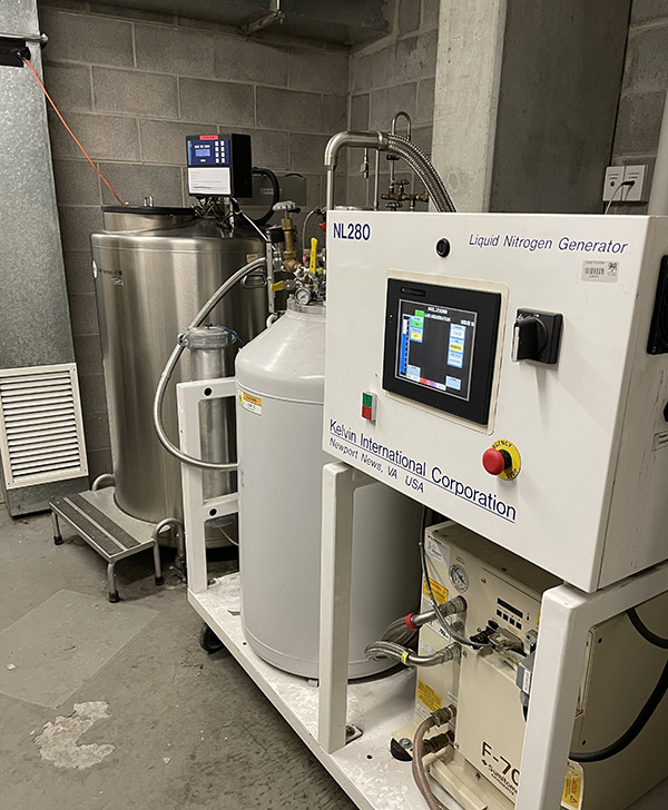
After shadowing Jo-Maree and the students in the lab, I feel pretty confident in moving forward with some actual cell work. We are just waiting on the delivery of new cell culture media. There is a bit of a delay with some orders due to the COVID lockdowns in NSW and VIC.

I did receive my glass Petri dishes (90mm and 1500mm) to do some initial tests and had a go at laser engraving them. I usually work in Perspex, so getting the settings just right is unfortunately always a bit of trial and error.

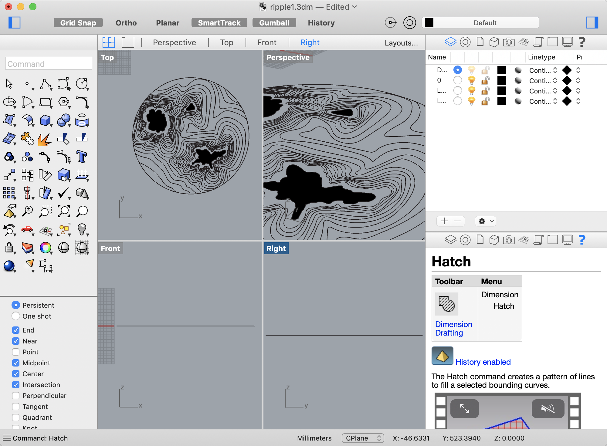
We decided to start with the simple ‘target’ engraving which would enable us to test different engraving depths and see if the graphic should use raster for hatched areas (circles) and vector for lines.

Graphic for Laser Engraving

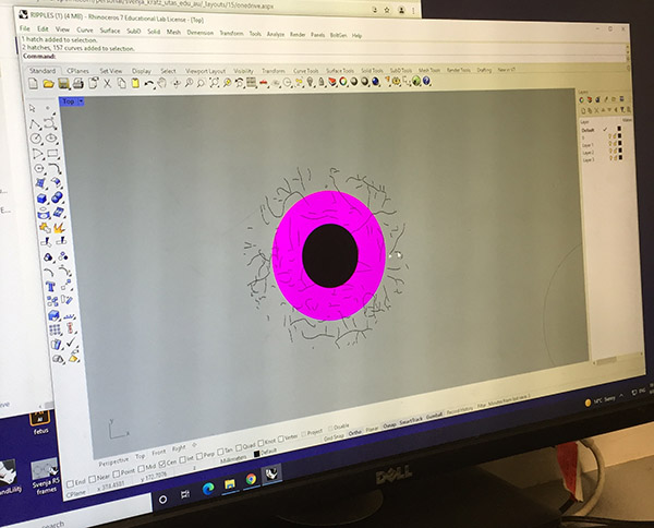
Graphic for laser engraving shown in Rhino 3D.

We need to leave room for the laser head to engrave so the graphic will be centred in the dish with a 25mm boundary. We also had to ensure that each element was on a separate layer with a colour indicating the particular settings for that graphic element.

We tested some basic settings:

Laser Engraving Settings

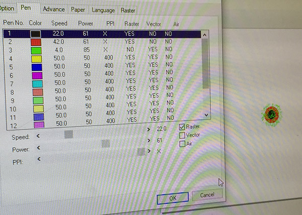
Black circle: Raster
Speed: 22
Power: 61
PPI: X
Red circle: Raster
Speed: 42
Power: 61
PP1: X
Green Lines: Vector
Speed: 4
Power: 85
PPI: X

We secured the sides with some metal rods to reduce the likelihood of movement when the air flow is turned on during the laser engraving process.

Laser Set Up
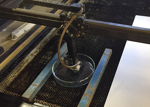
Set up in Laser  with metal bars to reduce movement. 
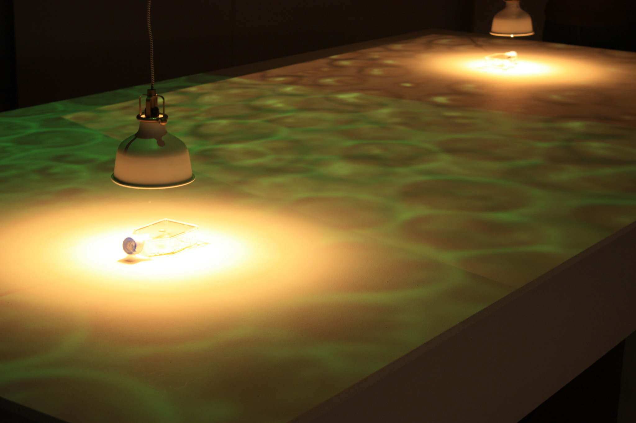
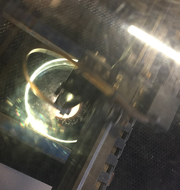
Laser Engraving ProcessLaser in process.

While the initial engraving seemed promising, the entire graphic was rastered including lines which were supposed to be rendered as vectors. The air flow also moved the dish. This resulted in an initial ‘glitch’ area and, following another shift, an overlaid section.

Engraving Glitch
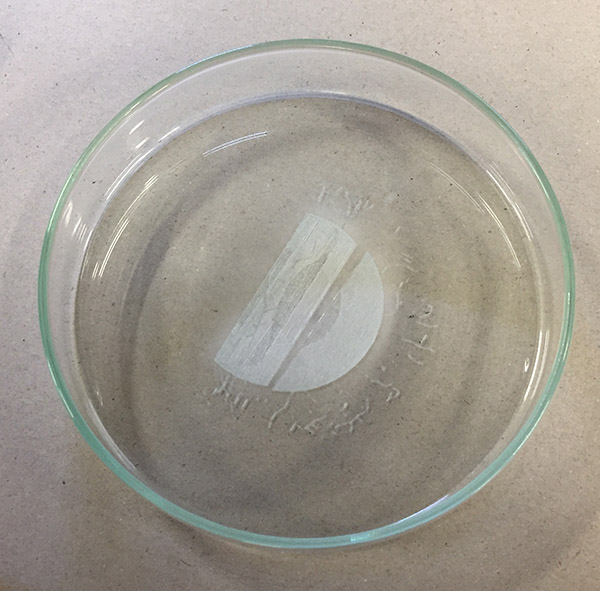
Laser engraved dish with ‘glitch’ pattern. 

It is not too bad, but shows the importance of testing settings and set up never assuming that things will work first time around.

We used the same dish again to test different settings (with four metal bars to hold the dish in place. However,  we found that the  raster setting was the best approach after all  to ensure that the laser creates lines without punching through the glass. We still don’t quite have the settings down, but hopefully the next go will yield more promising results.

Lab meetings including Science in the Pub Preview by Ash Russell

I have been attending weekly Tuesday Lab meetings at the Medical Sciences Precinct (MSP). This is a great way for me to gain insight into the research of group members. It is also wonderful to be immersed in the science lab culture where meetings become a platform to share work in progress and receive critical input from fellow HDR candidates and supervisors. These presentations usually focus on the research projects of particular  members – much of focuses on stroke research and is  is still in development/unpublished and therefore not suitable for sharing beyond the immediate group setting.

Science Meme

Science Meme via ‘Meme Your Science’ Event by Science in the Pub Tasmania Facebook Event

This Tuesday, was a bit of an exception as we got to listen to a preview of a public presentation by Ash  Russell,for the Science in the Pub Series in Tasmania this Thursday [5th August].  The theme is ‘memes’ so speakers have to use memes to convey their ideas. Ash is currently completing their PhD in bioinformatics, so the presentation focused (quite disturbingly) on the issue of scientific validity with reference to factors such as random chance and publication bias. The presentation was really entertaining so I do recommend heading along (noting that the face-to-face event has sold out, but you can stream along via the Facebook page.

Rhino Files

I have started preparing files in Rhino for laser engraving. This is a good Youtube introduction to setting up files with a some pointers for trimming and adding a hatch for engraving surfaces rather than just lines.

This is the start of my file prep in Rhino with dark areas signalling ‘hatched’ sections for surface engraving.

Screenshot of design in Rhino

Once the Petri dishes arrive, I can make time with the CAM technician Murray Antill to organise engraving. Of course, I need to test the settings first to ensure that I use the correct strength for lines vs. engraving. I usually use Perspex, so I will likely need to adapt the settings to suit a different material. The lines may also need further spacing as the laser produces around a .5mm line.

Immortalised cells – another work

As I continue to develop the project, I am looking into other artists working with cell immortalisation protocols.

A recent project involving the immortalisation of primary cells is the work Immortality for Two which forms part of a dual body of work collectively titled I’am by Luís Graça and Marta de Menezes. In contrast to the aims of the Billy Apple® project, this work reflects on the artist de Menezes and scientist Graça’s long-term working and romantic relationship (de Menezes M & Graça 2020).

Immortality for Two

Immortality for Two, Marta de Menezes 2014 – image available via Bioart Society

For more information see: https://martademenezes.com/portfolio/immortality-for-two/ 

de Menezes M & Graça L I’am – Immortality’s Anti-Marta, in Berger, E., Mäki-Reinikka, K., O’Reilly, K. and Sederholm, H., 2020. Art as we don’t know it. Aalto University School of Arts, Design and Architecture, pp. 52 – 53