Tag Archives: scaffold

Lockdown lingers…

Lockdown has lifted, but we have restrictions in place which limits access to lab areas unless absolutely necessary. Jo-Maree has kindly taken over caring duties and will pop in to feed my struggling fibroid cell colonies.

The HBVPs will be put to rest for now with scaffold tests fixed in 4% PFA. We may yet be able to stain them to determine if HBVPs were growing within the structure. Since the scaffolds are optimised for tissue/bone regeneration (and hence bone and tissue cells), they don’t seem to work too well with pericytes – so far anyway.

Since Jo-Maree had a stash of left over vials, we had planned to use Calcein to determine cell viability and visualise the cell growth along the scaffold structure as the scaffolds themselves seem to be non-fluorescent.

Calcein image via APB Biosciences
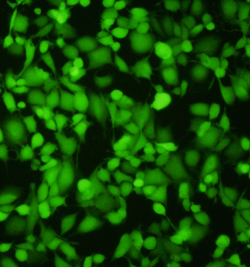
Impressive image of Calcein dye – live cells fluoresce a vibrant green – image via ABP Biosciences.

Since the Calcein dye works on live cells, we will need to reseed the scaffolds when lab-life returns to ‘normal’. This is fine as we will hopefully have enough fibroid cells by then to use for the scaffolds and also undertake fluorescent microscopy – i.e. use antibodies to reveal cell cytoskeleton details (e.g. actin filaments) and DAPI  blue-fluorescent dye for nuclei.

Fluorescently labelled cell via Leica
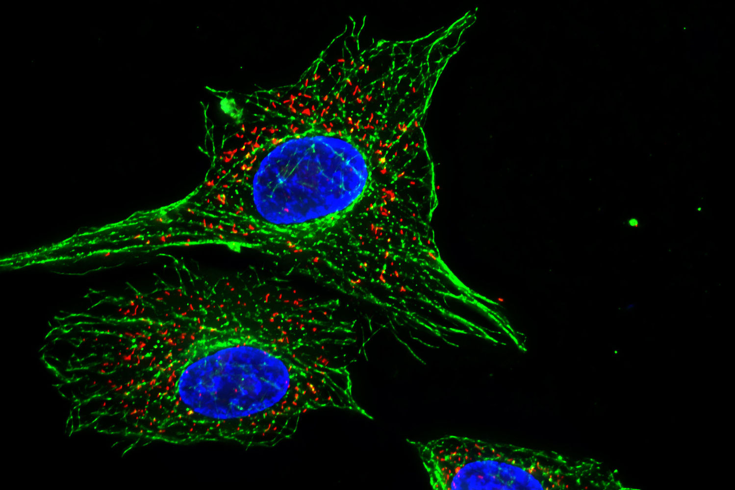
Image of fluorescent cells via Leica. 

Sponges as scaffolds?

We are lucky in Tasmania to be able to travel freely across the state. With winter coming to an end, I saw an opportunity to visit Burnie with some art school colleagues. We witnessed the arrival of some of the first penguins at the Burnie Little Penguin colony for their annual mating and childrearing.

We also glanced some amazing sponges on the beaches in nearby Wynyard.

Wynyard BeachFossil Bluff – Wynyard.

Doctor's RocksDoctor’s Rocks – Wynyard

Marine Sponge texture

Seeing the texture and architecture of different marine sponges on the beach, prompted me to consider whether they have been considered as a scaffold architecture for cell growth.

Sponges from Wikimedia
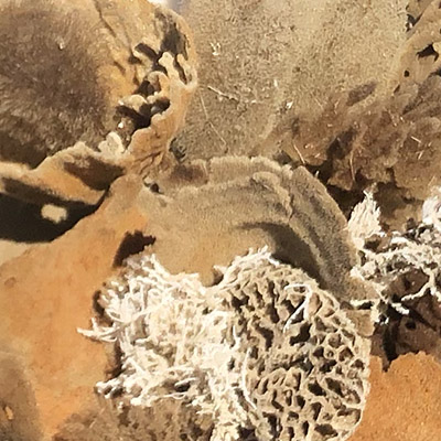
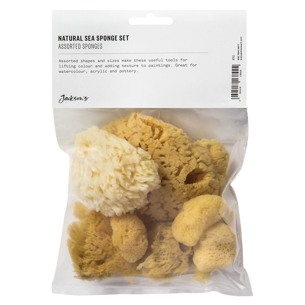
Different sponges from Wikimedia Commons.

Turns out that yes, there is already a study on whether marine sponges could be used as scaffolds in bone repair.

In vitro Evaluation of Natural Marine Sponge Collagen as a Scaffold for Bone Tissue Engineering

While this has already been done. I think it would still be a nice side experiment to see whether I can grow my cells in a marine sponge scaffold. There are a number of companies that offer cleaned and bleached natural marine sponges for bathing, facial exfoliation and art – although the variety seems usually limited to honeycomb and silk sponges from the Mediterranean.

Sponges

Bag of sponges available from art supply store.

Perhaps the sponges could be used in conjunction with a hydrogel to assist with cell adhesion and proliferation. I think it would be quite lovely to make a self-portrait of ‘me’ as a sponge. Although, I will likely need to use a bioreactor to enable nutrients to reach the interior of the structure.

Scaffolds Arrived

Dietmar’s scaffolds have arrived.

Scaffolds in Express Post BagScaffolds arrived via Express Post on 16/09/21

This means that we can do some tests to see how the HBVPs grow in a matrix.  The scaffolds include some flat and tubular sections for initial experimentation.

MPC Scaffolds
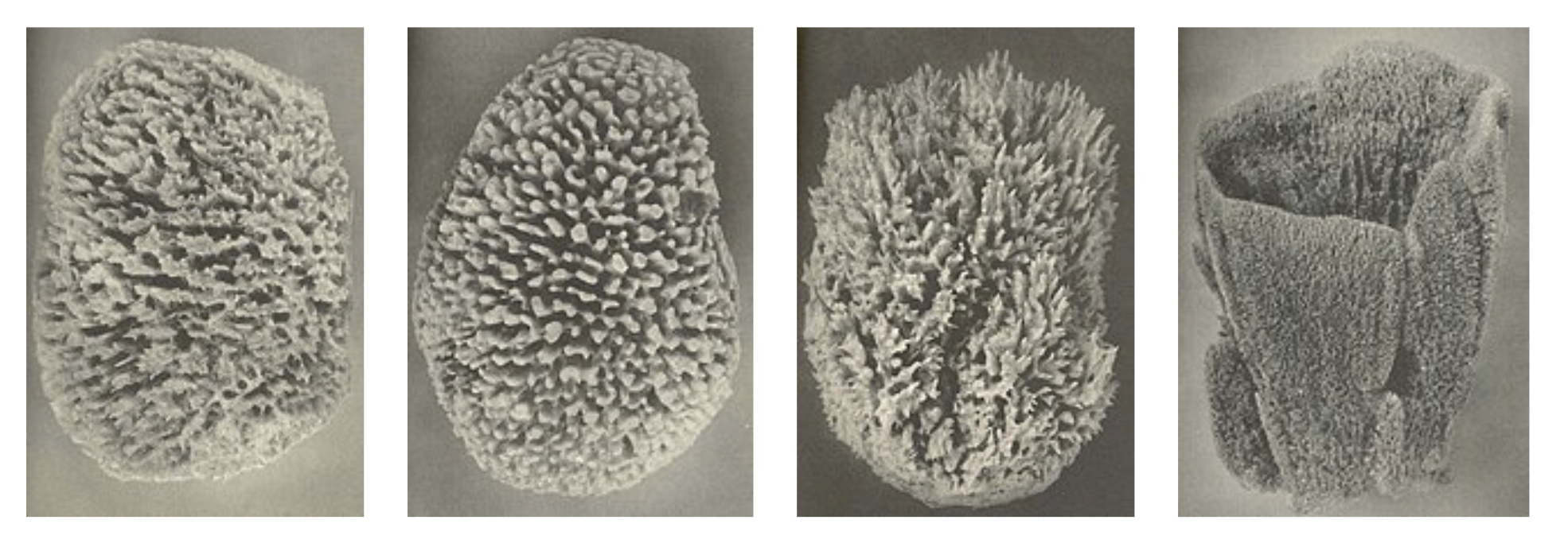
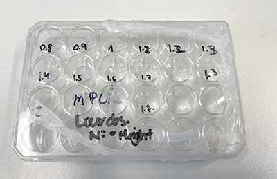
MPCL Scaffolds – tubular structures at varying heights.

Scaffolds in Petri Dish 
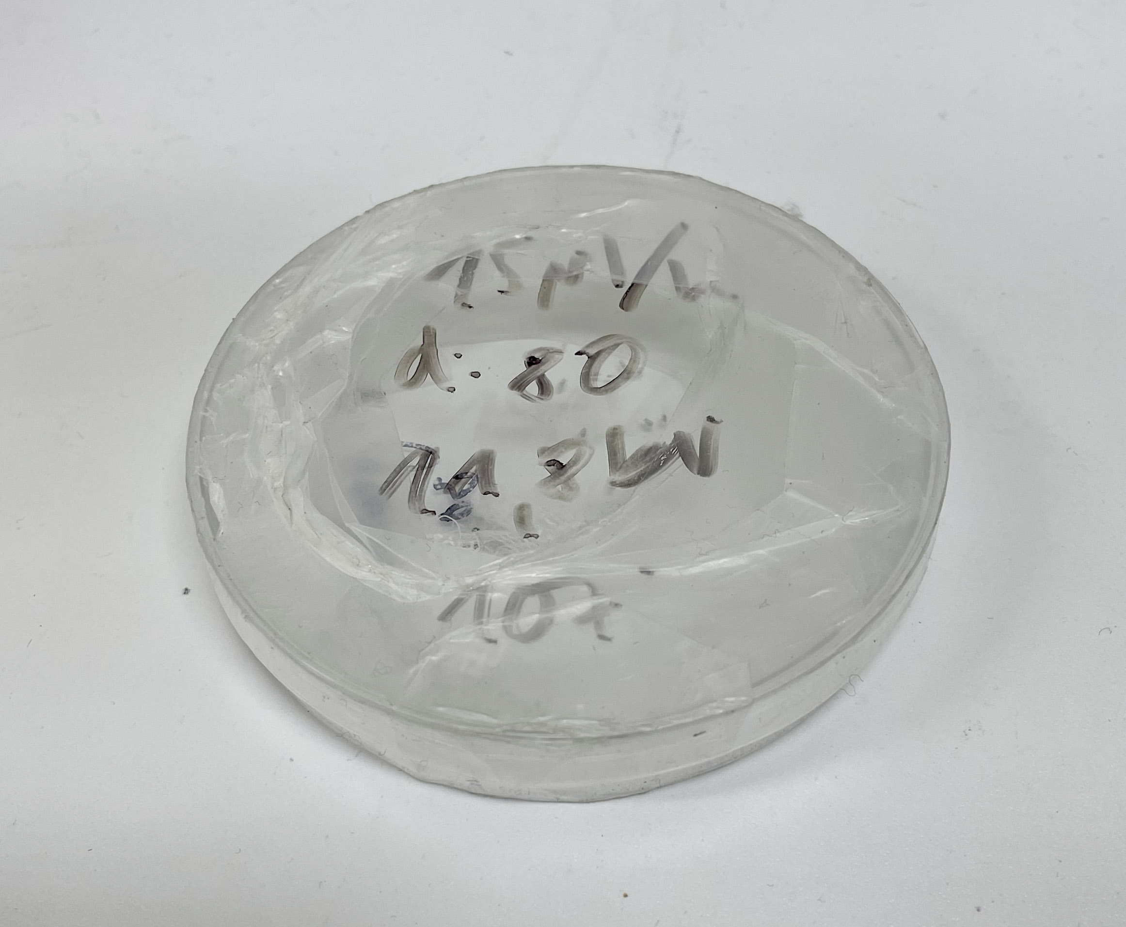
Scaffolds in Petri Dish  – flat square structures. 
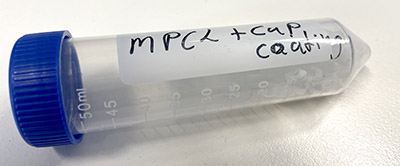
I will need to confirm with Dietmar’s group but the notations seem to indicate that the scaffolds are  medical grade polycaprolactone (mPCL) with some tubes including a  calcium phosphate (CaP) coating.

MPCL+CaP CoatingMPCL+CaP coating

MPCL+CaP Coating
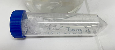
MPCL+CaP coating – tube structures.