Preparing the ‘Dirty’ Lab

We are almost ready to culture my little tumour baby cells. Since they are primary cells, we need to work in the ‘Dirty’ Lab area. As previously mentioned, this does not mean that the lab is any less clean or sterile, but rather relates to the type of cells used. Since my fibroid cells are primary cells and untested for mycoplasma and other potentially infectious agents, they cannot be cultured in stem cell and cell line areas.

'Dirty' Room

“Dirty” Cell Culture Lab

The incubator has been out of action, so the first task is to clean the shelves, change the sterile water and decontaminate the interior.

cleaning

Washing the incubator shelves

The incubator we are using has an auto-clean function that super heats the interior to sterilise everything. A nice feature!

incubator

Incubator

Once the incubator has gone through the full cycle (approx. 24 hours), it will be ready to host my cells.

Using the Autoclave

Jo-Maree inducted me into the use of the benchtop autoclave used to sterilise glassware and other consumables (e.g. water) for use in the lab.

Benchtop Autoclave

I was a bit nervous at first, but the actual process is pretty straightforward with clear instructions on the front of the machine.

Autoclave Instructions

The cycle setting depends on the nature of items that need to be sterilised. I will be autoclaving Glass Petri Dishes in autoclave bags. While there is no specific ‘Goods’ listing for glass, Jo-Maree indicated that Cycle 6 – Wrapped Metals – would be most appropriate as the glass can withstand high temperature and is comparable to metal.

I will also need to sterilise some MilliQ Water for cell culture use – e.g. washing dishes, so this would need to go under a different cycle – Cycle 2 for fluids. When sterilising fluids, it is important to only loosely add a cap to the vessel or the bottle can explode.

Clay surface tests

Alongside 3D sculpture tests, I have also been working on some old canvases to see how the chameleon pigments work on a more 2D surface. These canvases have hand mould clay elements that resemble worms and bacteria. I must admit that I am not quite sure where I am going with this… although at the very least, it will enable me to build up layers of chameleon and UV reactive pigments.

Clay Panel

Canvas with 3D clay surface texture and first coating of chameleon pigment.

Chameleon Pigment: 3D Experiments

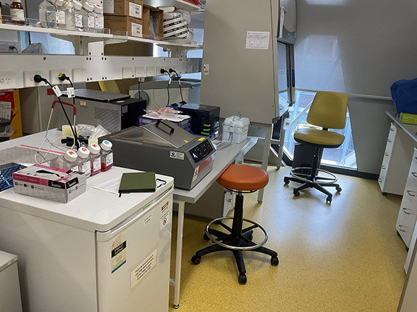
While I have been prepping for lab work (and awaiting clearances and media), I have been using my residency to invest in studio time and test new materials and processes that (somewhat subconsciously still) align to the themes of the Synapse project.

Over the past few months, I have ordered a stack of chameleon pigments from suppliers in Australia, China, the US and UK to test quality and colour. I have compiled a series of swatches on different substrates to determine colour shifting properties and how they layer and complement each other. So far my favourite supplier is A1 Pigments. They stock a huge range of powdered pigments including chameleon and mica pigments.

I have also tested solvents including water-based varnishes, oil-based media and wood varnish. Unfortunately water-based products are not suitable for the pigment and it works best in a polyurethane/resin base. I particularly like the results from using timber varnishes such as the Bondall Monocel Timber Varnish range. They have a slight warm amber tint which  gives the application a nice depth of colour. For crystal clear application and sealing, KBS’s Diamond Finish Clear Coat is fantastic as it is non-yellowing and super clear (although a bit expensive).

As part of my testing process, I have started to develop a series of small sculptural works that consider species and matter entanglements and the wonder of the universe.

Sculpture 1

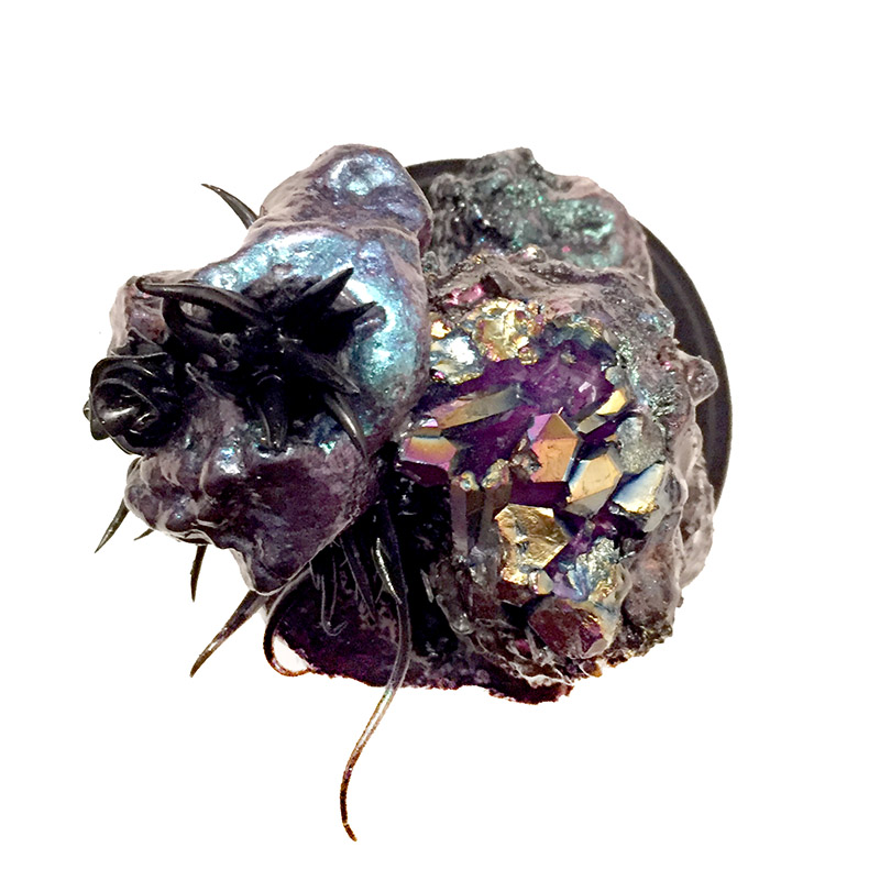
Top view of Sculpture 1 showing chameleon pigment coated rocks formations and Titanium Aura Quartz

Sculpture 1

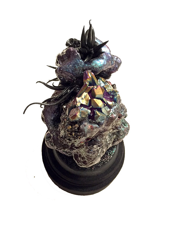
Frontal view of Sculpture 1

Sculpture 2

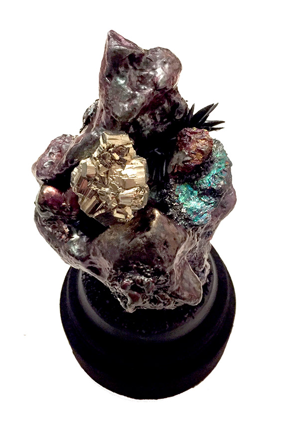
Frontal view of Sculpture 2 showing a Pyrite cluster and Peacock Ore (Bornite).

These sculptures have ‘secrets of the universe’ hidden in their base. The form of the works hints at the nature of the secret, but you would have to destroy the work to reveal it.

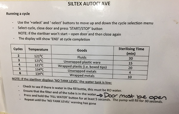
I really wanted the base to be a dark matt black. To achieve this I tested Stuart Semple’s Black 3.0. While the initial results were good, the black marked too easily with my messy handling. As such, I opted for black flock  instead. Not only does this coating hide slight surface imperfections, it also creates a great matt black and light absorbing coating. Now that I am making some headway on ideal flocking application, I am planning to flock some of sculptural works to create a contrast between soft velvety textures and glossy surfaces.

I should mention that was first introduced to flocking by my dear friend and fellow artist Michael Riddle. Finally, I have a purpose for my own work to ‘get flocked’. So thanks Mike.


Sending mouse sperm via postcard

The great thing about residencies is that you get to spend a good amount of time talking and sharing stories and insights. At our lab meeting, our conversation led to this awesome paper:

Mailing viable mouse freeze-dried spermatozoa on postcards

Yes, you read correctly.  It is indeed possible to share mouse sperm via postcard.

My favourite part are the graphics:

Mouse Sperm

Thanks Jo-Maree 😉 I think this is another potential project…

LAB SCHEDULE

UPDATED LAB SCHEDULE

Now that the project has the formal go-ahead, I am moving into lab mode and have determined some of the key milestones for the next months.

1: Training & Prep: 1 – 2 weeks

Training with HBVP cells include:

  • Thawing and culturing cells, making media, working in a biosafety cabinet and maintaining sterility, light microscopy
  • Learn to use the autoclave and prepare petri dishes and glass vessels for culture
  • Coat petri dishes and glass vessels with poly-l-lysine for cell adhesion, test with HBVP cells
  • Order media, reagents and kits
  • Submit IBC approval forms

2: Cell culture of fibroid cells – 4 – 8 weeks

  • Thawing and culture – grow up and freeze stocks of cells, light microscopy
  • Ask Dietmar to send 3D scaffolds
  • Grow and fix cells in petri dishes and glass vessels
  • Fluorescent microscopy of cells
  • Scanning Electron Microscopy (SEM) of cultured cells
  • Transmission Electron Microscopy (TEM) of cultured cells
  • Timelapse microscopy
  • 3D cell seeding HBVPs and Fibroid cells – see differences in cell response.
  • Wait for IBC approval

PROJECT: 3 months

3: Cell Immortalisation +

  • Immortalisation of primary fibroid cells via established commercial kit (Applied Biological Materials (ABM) or Alstem cell immortalisation kits)
  • Cell genetic profiling
  • Cell culture of immortalised fibroid cells (optimisation of culture methods for 2D and 3D environments, cellular response and proliferation testing)
  • Grow and fix cells in petri dishes and glass vessels
  • Timelapse microscopy

4: iPSC production

  • Reprogramming of primary cells to generate induced pluripotent stem cells (iPSCs) via established commercial kit (e.g. Epi5™ Episomal iPSC Reprogramming Kit available via Thermo Fisher)
  • Development of Gastruloids, Organoids or Neurospheres (self-organised 3D cell masses)
  • Cell culture of reprogrammed or immortalised fibroid cells (optimisation of culture methods for 2D and 3D environments, cellular response and proliferation testing)
  • Timelapse microscopy
  • If iPSC successful – create neurons and heart cells

Ethics GRANTED! …but one more approval to go…

We have approval to move forward with the key aims of the project. This is great news as it means I can start working with own cells. I am still a bit precious as there are limited vials, so I will do a couple of weeks of training on HBVPs before I move on to my own cells.

While we can get started on the fibroid cell culture, Brad realised that cell immortalisation will require further Institutional Biosafety Committee (IBC) approval. This is because the process will require the uses of lentiviral vectors. As such, it is considered Notifiable Low Risk Dealing (NLRD) and the committee will need to ensure that we have the appropriate facilities and training in place to move forward.  iPSC cell reprogramming is exempt, but we still need to let the IBC know what we are doing.

The application is due tomorrow, so we had a meeting this morning to go over the protocols and identify the particular kits we are going to use. There are a range of biomedical research supply companies, but the important thing is to make sure that we use an organisation that has an Australian supplier.  hTERT and SV40 T Antigen kits are the best options for our project as they are suitable for a range of  cell types including fibroblasts.  Fischer Scientific have Alstem Immortalisation Kits available, but ABM may also be a good option. They also have a good overview of Cell Immortalisation Protocols for anyone interested in the process.

For iPSC reprogramming, we are going to use the Epi5™ Episomal iPSC Reprogramming Kit by Thermo Fischer. Another group has used this product previously – so we can get tips on how to get the best results.  Lovely Jo-Maree is looking into the best purchasing options. With lead time for purchase and delivery, the products will likely arrive around the same time as final approval.

Cell Immortalisation Products

In preparation for meeting with Brad and Jo-Maree, I compiled a list of cell immortalisation products. Many of the companies listed are US, so this may impact on availability.

CELL IMMORTALIZATION KITS:

  • ALSTEMBIO: SV40 T Antigen Cell Immortalization Kit 

GENTARGET: SV40 Large T antigen  

GENECOPEIA: Cell Immortalization Reagents 

FISCHER SCIENTIFIC: Alstembio 

CAPITAL BIOSCIENCES: Lentiviruses for Cell immortalisation 

ABM: Cell Immortalisation 

BIOCAT 

CREATIVE BIOARRAY 

Exhibition Planned for June 2022: Preliminary Creative Work Ideas

I am excited to more formally announce that I have an exhibition scheduled for June/July 2022 at The Barracks Gallery in New Norfolk, run by Derwent Valley Arts. This is a great opportunity to show preliminary outcomes from the Synapse residency. A deadline also always gets me moving creatively. Due to the heritage location,  I am not anticipating showing living works, but rather fixed cells as part of sculptural works and other mixed media works and prototypes.

While final creative works will of course be refined in response to laboratory outcomes and collaborator input, these are some preliminary ideas:

  • Tumour Babies: A series of six media-media wall panels integrating my DNA and stained and fixed cells grown in glass vessels.
  • Revelations: A series of six mixed media dome works incorporating fixed cells seeded into 3D biofabricated scaffold structures.
  • More-than-human: A large-scale sonic and LED chandelier integrating 3D printed resin components based on cells, microorganisms, protein and DNA structures
  • Visible/Invisible: A series of three UV activated paintings and laser engraved light panels
  • Self-portrait #4: Imminent unfoldings: An outdoor sculpture designed to transform in response to the environment.
  • Becoming (M)other: Sculptural video installation integrating time-lapse light and fluorescence microscope images of cell growth and transformation.
  • Legal Mandalas: A series of laser engraved mandalas incorporating text from relevant legal and governance frameworks related to biomaterials use and patenting in Australia over the past decade.


iPSC Protocols

We are still waiting for formal ethical clearance to undertake work using my fibroid (fibroblast) cells. In the interim, Brad sent through a Nature protocol detailing options for producing iPSCs from human keratinocytes derived from plucked hair follicles or skin biopsies. I have isolated keratinocytes from hair follicles before at QUT when I was part of the Tissue Repair and Regeneration Group for  the HSE (Human Skin Equivalent/Experience Project).  It was quite a mission as keratinocytes require a feeder layer of fibroblast cells. These needed to be irradiated to ensure that they did not outgrow the keratinocytes 🙁

Since we are only doing a skin biopsy if the fibroid cells are not viable, I am parking this option and scouting for protocols that are fibroblast specific. With that in mind, the company Sigma-Aldrich has specified an iPSC reprogramming protocol – Reprogramming of Human Fibroblasts using Non-Integrating Self-Replicating RNA Vectors – designed for fibroblasts and with a 30-day creation estimate. Of course, you always have to double or triple timeframes when you are undertaking a protocol for the first time. Of course, I will need to discuss this option with Brad and seek a pricing and availability estimate.

Fisher Scientific also has a reprogramming kit – the CytoTune-iPS Sendai Reprogramming kit. However, the latest version and full kit carries a hefty price tag at over $18, 500. They also offer a potentially more affordable option via ‘Episomal Vectors’ or Epi5™ Episomal iPSC Reprogramming Kit. These could be an option but Brad is of course the best advisor.

There are also a number of journal articles detailing iPSC reprogramming including: Reprogramming fibroblasts into induced pluripotent stem cells with Bmi1 [Nature], Human Pluripotent Stem Cells (iPSC) Generation, Culture, and Differentiation to Lung Progenitor Cells [Methods Mol Biol.], Guidelines and Techniques for the Generation of Induced Pluripotent Stem Cells [Science Direct], Generation of human iPSCs from cells of fibroblastic and epithelial origin by means of the oriP/EBNA-1 episomal reprogramming system [Stem Cell Research & Therapy].

Looks like I’ve got some reading to do…