Since the cells showed the first signs of attachment on the 20th of April [Day 8], I monitored the plates on a daily basis to see the emergence of more reprogrammed PBMC (R-PBMC) colonies (precursor iPSCs) forming on the base of the culture dish.
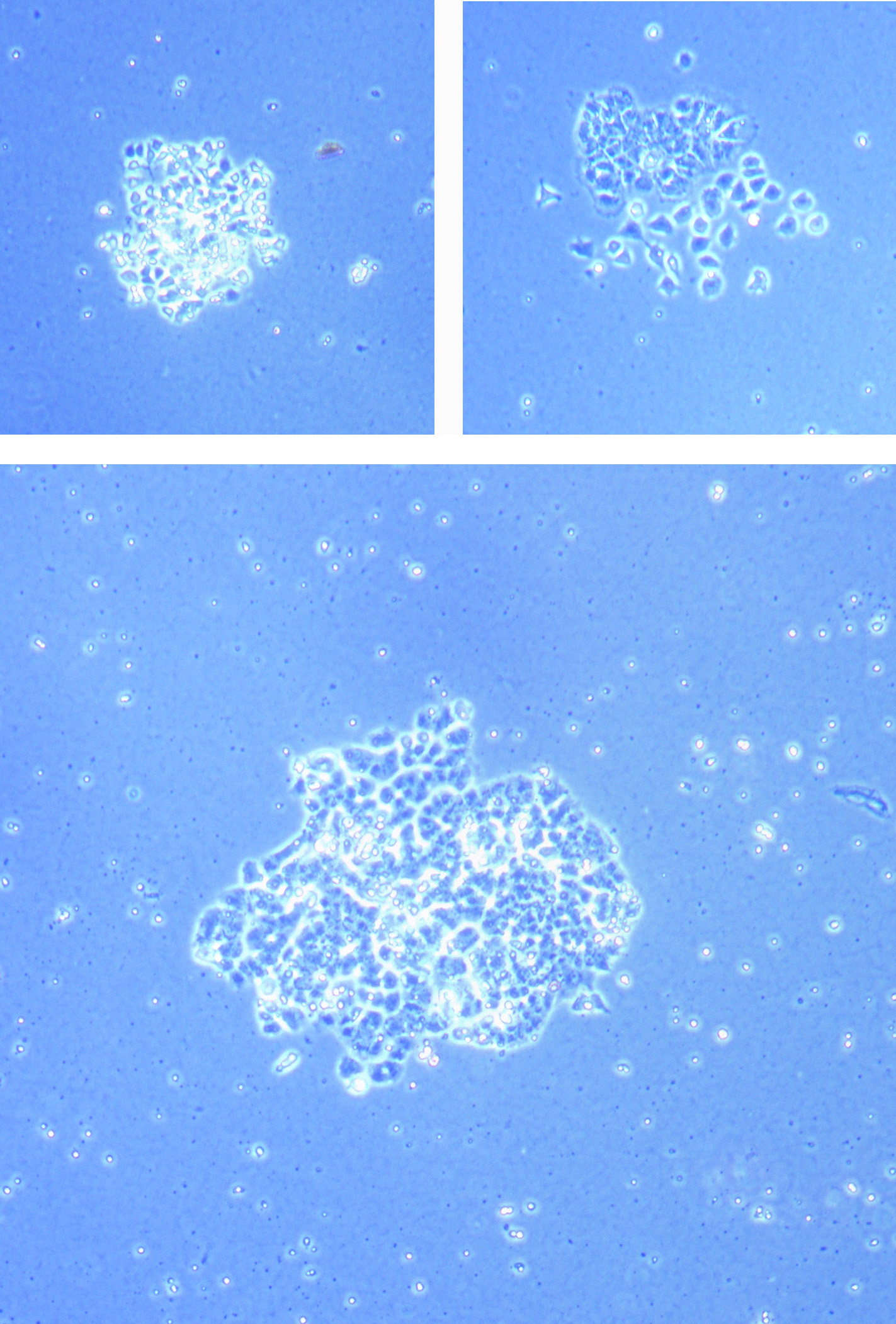
Plate #2 had colonies on the 20th, so there were some great looking cell clusters visible a couple of days later on the 22/04/22.
Good series of R-PBMC (precursor iPSCs) visible in Petri Dish #2 – 22/04/22
Petri Dish #1 was slower for colonies to emerge. However, by the 22nd of April, there were a couple of attached cell clumps .
A couple of R-PBMC (precursor iPSCs) colonies visible in Petri Dish #2 on 22/04/22
By the 24/04/22, the initial adherent cells were starting to proliferate well. While attachment and cell growth of any kind is always a good sign, we were keenly hoping to see the emerge of iPSC-like cells. These tend to clump together into small circular clusters.
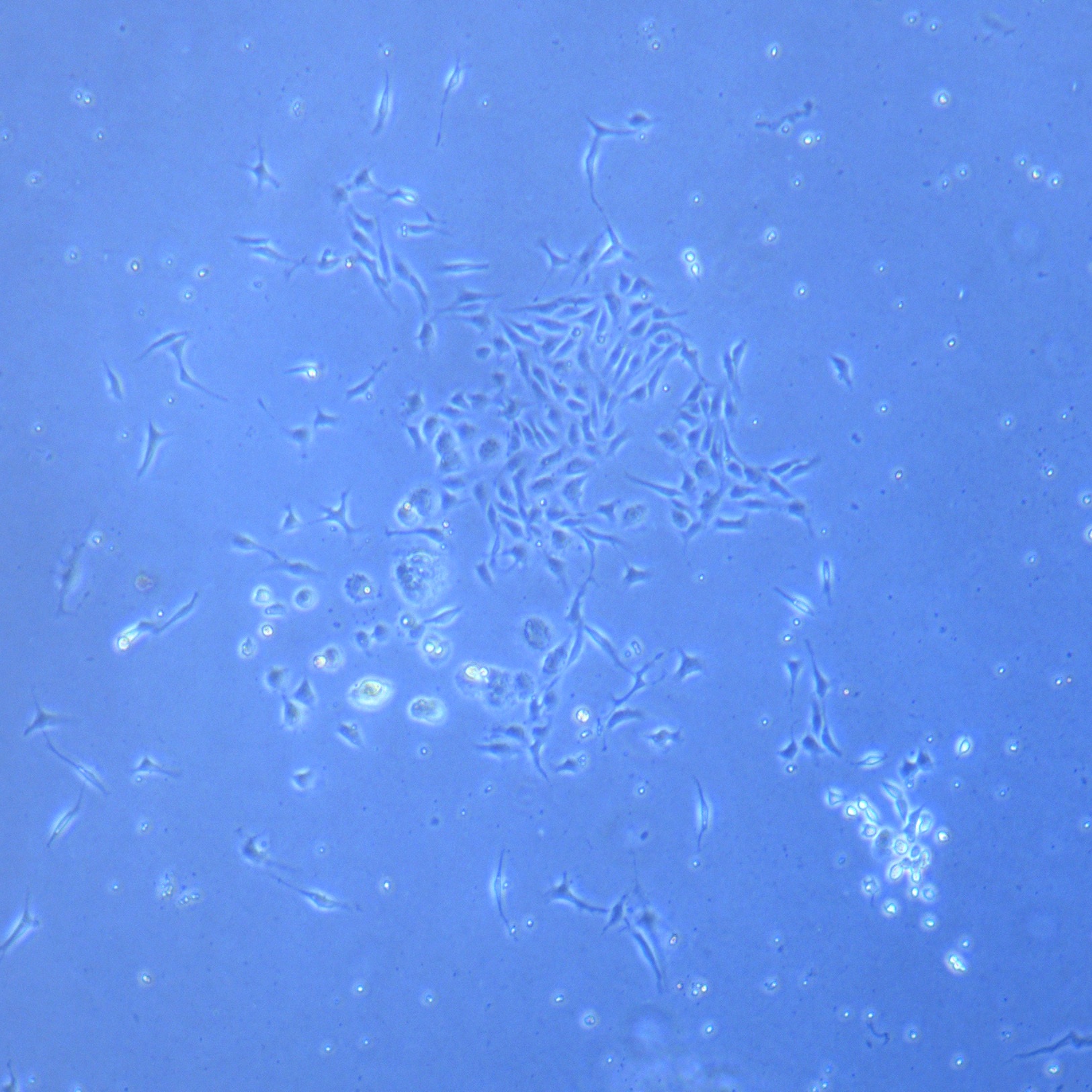
Attached cells with non-iPSC-like morphology on 24/04/22 in Dish #2Cells with a more promising iPSC-like morphology in Dish #2 on 24/04/22
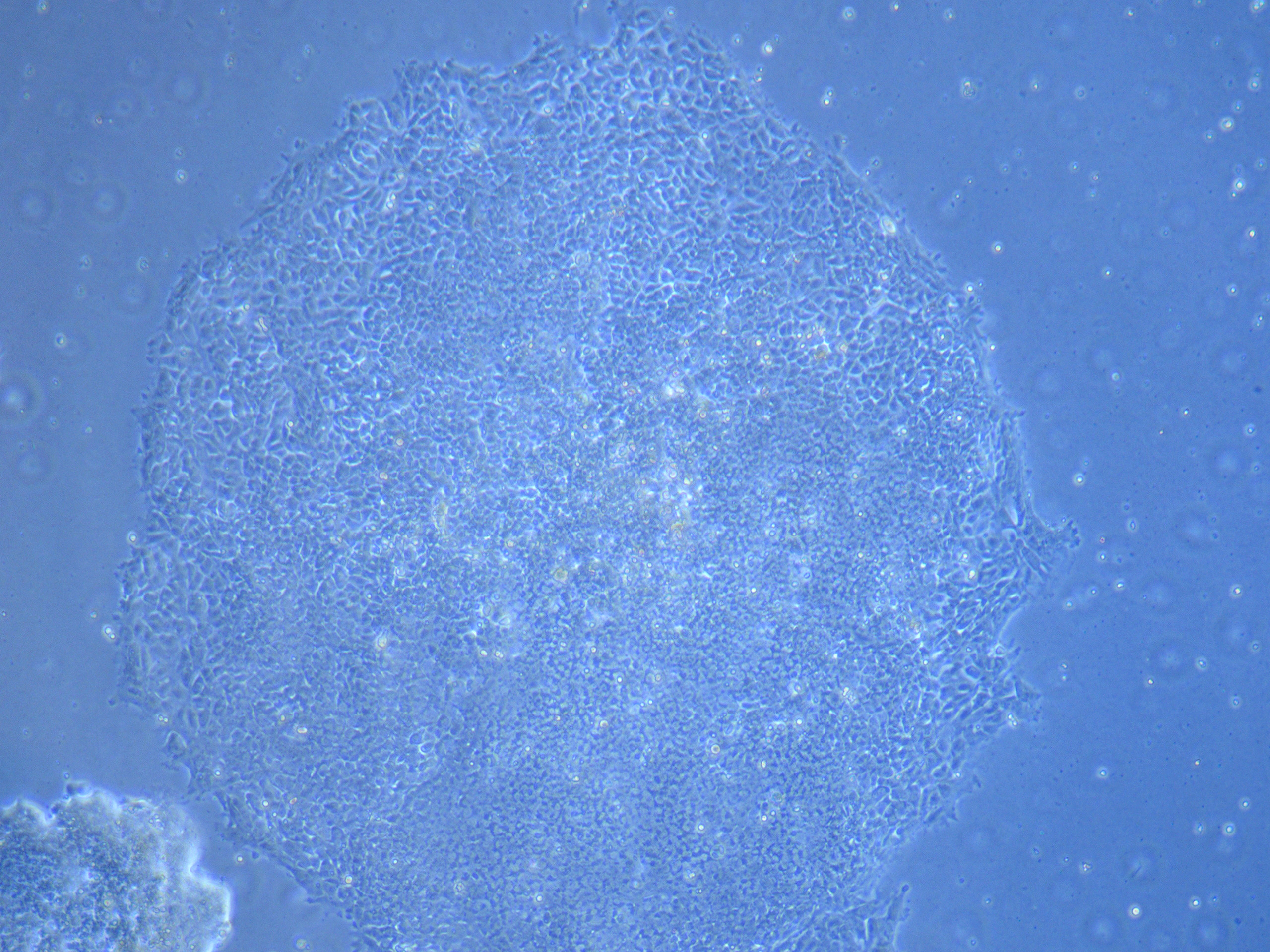
At this point the cells were still maintained in PBMC transition media, but by the 27/04/22, plates were looking good and we started to shift them to iPSC media. By 28/04/22 [Day 16], the cells are almost able to be classified as iPSCs.
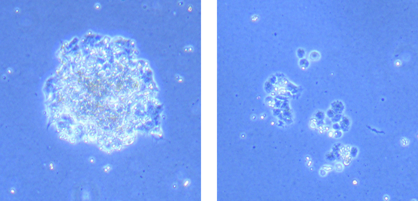
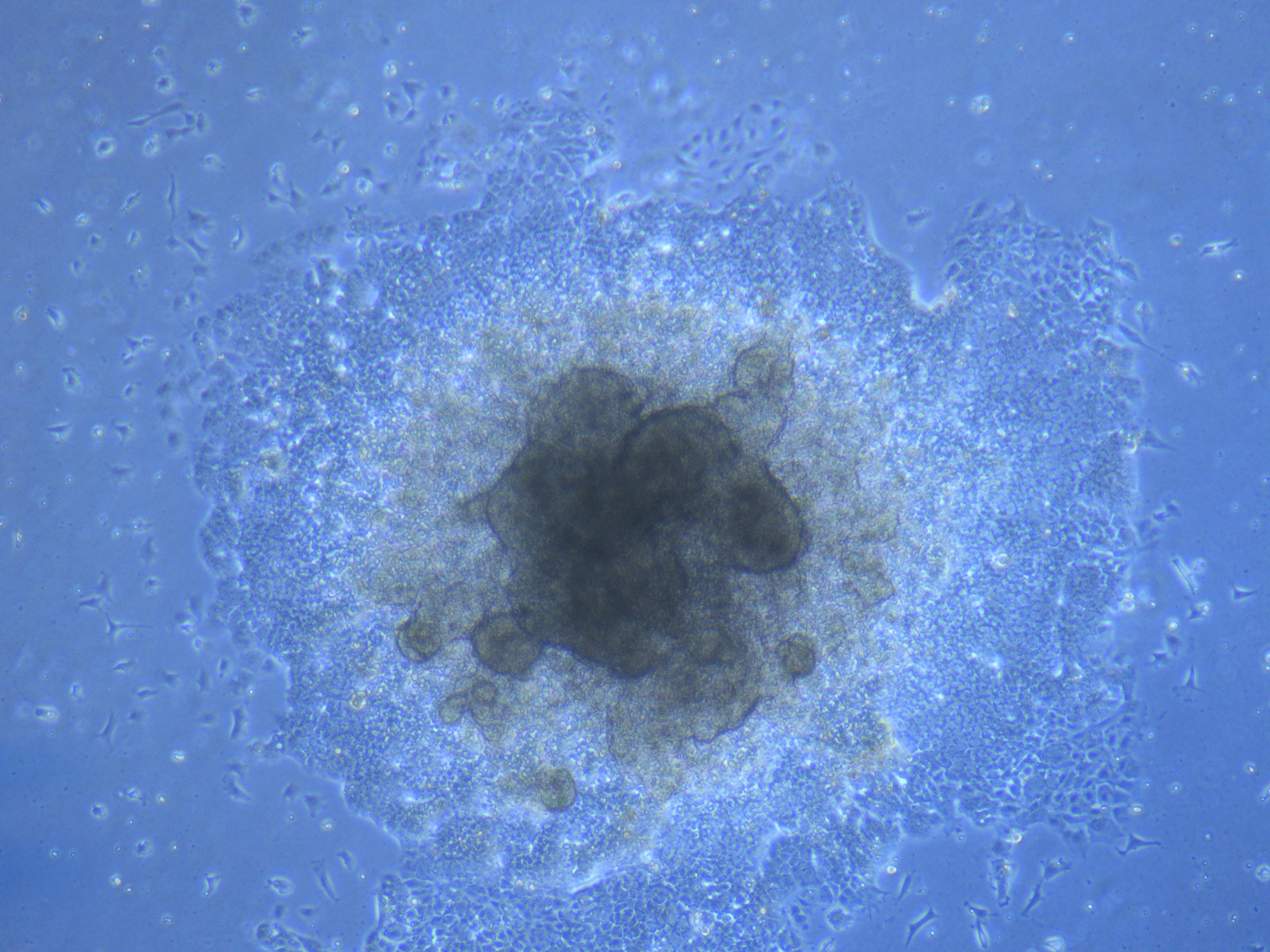
Large mostly uniform colony of precursor iPSCs on 28/04/22.More ‘yucky’ colony of precursor iPSCs with cell differentiation visible along the edges of the colony on 28/04/22.
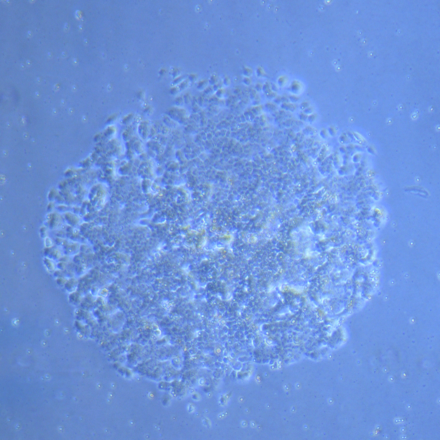
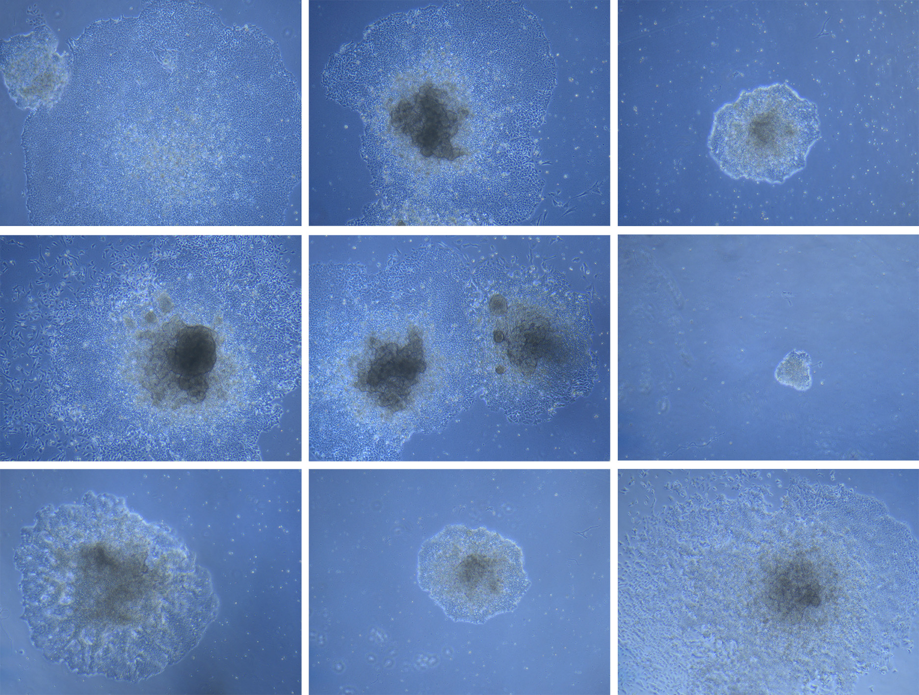
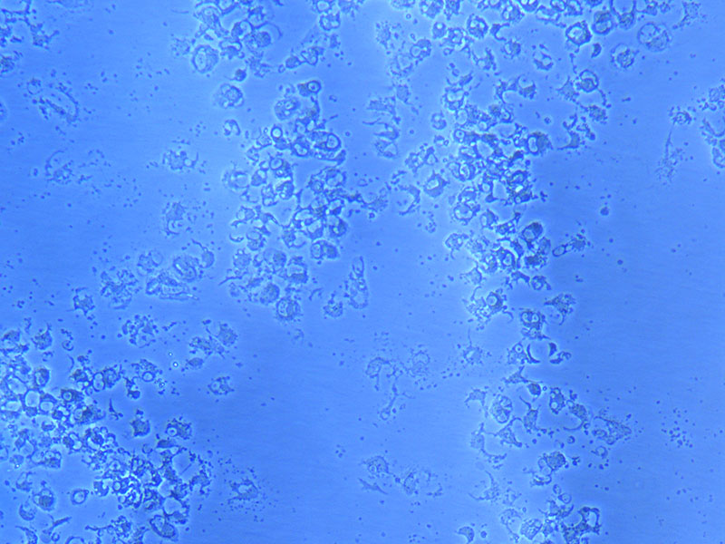
A day later on 29/04/22, the colonies were well and truly growing with a mix of large and small colonies (and some undesirable cells types).
When we checked our dishes on the 20th April, we noted that one Petri dish (Dish #2) has definite attachment. This means that we can start transitioning to new media. The other dish (Dish #1) is lagging behind. This is not necessarily a bad thing, but it will delay the media transition for a couple of days. Hopefully the extra time will result in beautifully formed colonies 🙂
Dish #2 has attachment – i.e. small colonies of attached cells starting to form on the base of the Petri Dish.
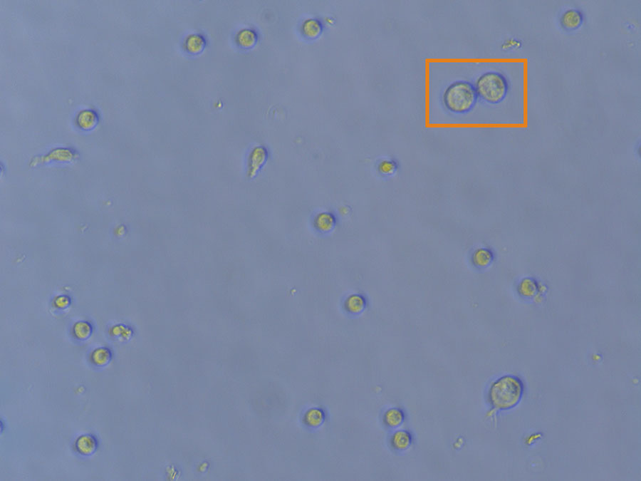
Observing the cells on 13/4/22 (24 hours after the transduction), Ash was confident that the virus had worked. While not scientifically verifiable, one of the potential indicators for success is the difference in size between standard PBMCs and ‘bloated’ PBMCs that suggest the cells have taken up the virus.
Microscope image of cells after transduction on 13/4/22. Larger cells indicating viral uptake are outlined in orange.
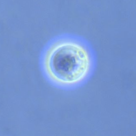
In addition to being larger, cells that may have taken on a viral load tend to have a speckled appearance. The bright halo also indicates that the cell is alive.
Microscope image of a large and speckled cell indicating that it has likely taken up the virus. Over the next few days, this cell may start to change and attach to the base of the culture vessel.
We transferred the cells to 2 x 60mm Petri Dishes coated with Matrigel. At this point, we just need to top up the cells with fresh media and wait (and hope) for transformation and attachment. We can also refer to the cells as RP (reprogrammed cells) rather than PBMCs.
Despite low PBMC cell numbers, due to my ineffective media change, we were able to proceed with the virus addition as scheduled. Ash is confident that we will still get a few colonies.
First we transferred the PBMCs to a new 24-well plate with fresh media. The reprogramming process, as outlined previously, is deceptively simple and involved adding a specific volume of each viral vector (total of 3 vectors) to the PBMC culture. Thankfully, Ash calculated the miniscule amounts required.
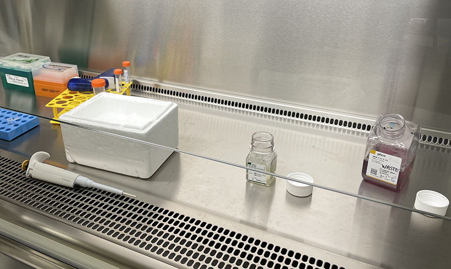
The virus must be kept cold. As such, the 3 vials were collected and kept on ice during the transduction process.
Virus vials on ice and in the biosafety hood ready for transduction of PBMCs
After the virus was added, the PBMC culture was placed back in the incubator for 24 hours.
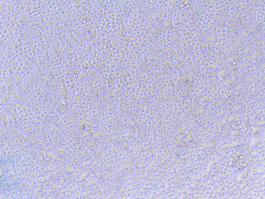
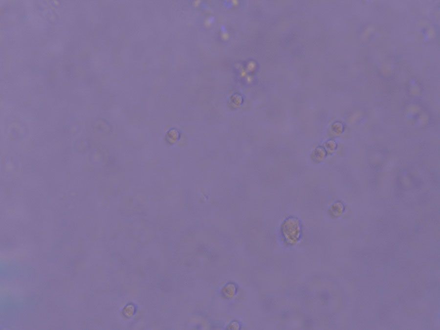
On Sunday 10th April, I did another media change for the PBMCs. The number of cells is definitely reduced. When I checked the previous well, there were a number of dead cells visible, so I have clearly left a lot of cells behind.
Light microscope image showing evidence of dead cells in the previous well. This illustrates that I was very successful at transferring all cells during the media change.
This shows the importance of mixing the cells well and checking that most cells have been transferred during the media change process. I will need to be really careful and follow Ariane’s instructions carefully to avoid further cell losses.
At this stage, we only have around 1.7 x 10⁵ cells (according to the cell count)!
Light microscope image of PBMC Cells taken on 10/4/22
On the positive side, there is no evidence of contamination and apart from low numbers, the cells seem healthy.
As per our scheduled timeline, we performed the first PBMC media change. This involved collecting and spinning the cells, then resuspending them in fresh media. While the process is quite straightforward, it is tricky working in a small well. I have a feeling that I did not mix well enough and may have left too many cells behind in the well.
PBMC Cells on 8/4/22 prior to media changePBMCs after media change on 8/4/22
There is a remarkable difference between the before vs. after photos, even though it is difficult to photograph cells in suspension just after they’ve been passaged. We will see how they are faring tomorrow…
At least, the frozen PBMCs are safely stored away in liquid nitrogen now!
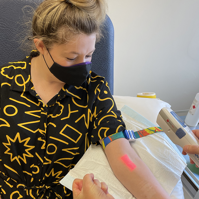
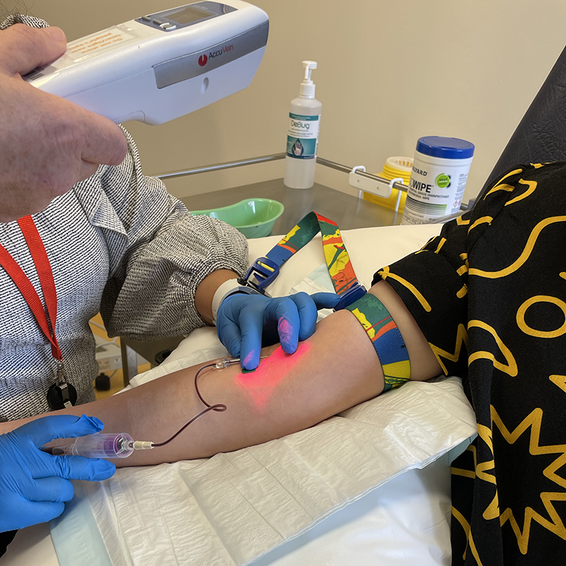
At the UTAS Medical Science Precinct, we are fortunate to have experienced phlebotomists on site to undertake the blood collection process.
Blood Collection
We started the collection process at around 9:30am. My first vein (left) was difficult to find, even with the use of a fancy vein illumination machine. However, my second arm (right) had a good vein for blood collection resulting in a 5mL sample for processing.
Image documenting first attempt to find a viable vein for blood collection.Image documenting successful collection of blood from right arm.
PBMC isolation
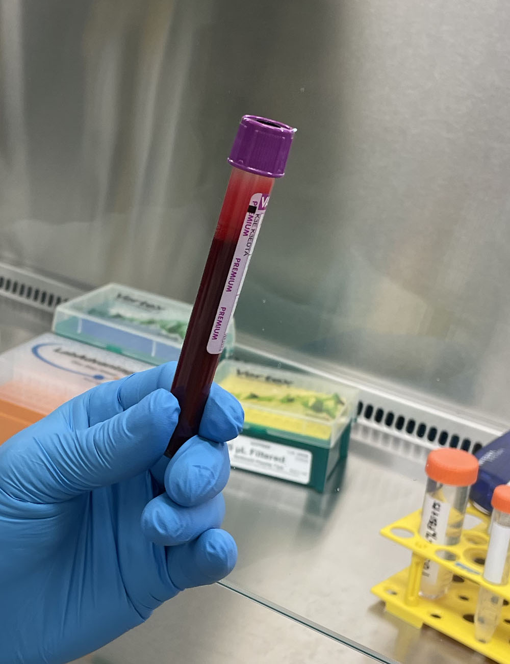
Blood sample in lab.
We transferred the blood to the lab and processed the sample straight away to obtain PBMCs.
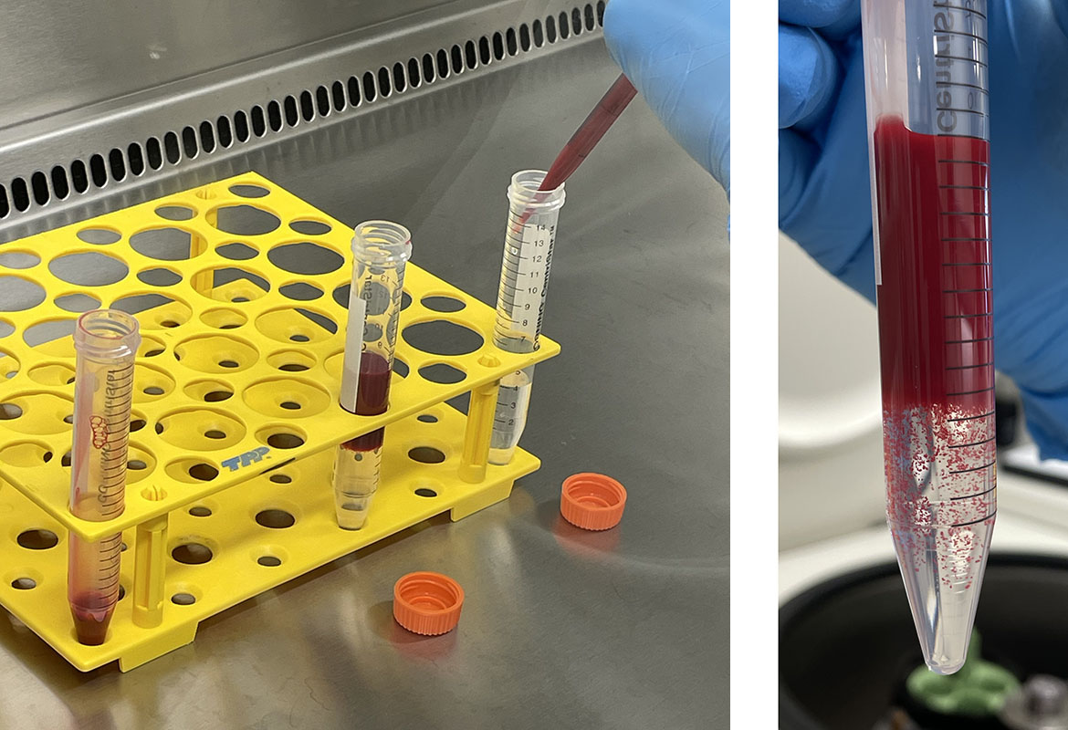
The first step involved diluting the blood sample with PBS (saline solution) to make it less viscous. This was then gently added to a tube containing Ficoll solution to form a clear layer of blood over the medium.
Blood added to gradient medium ready for centrifugation.
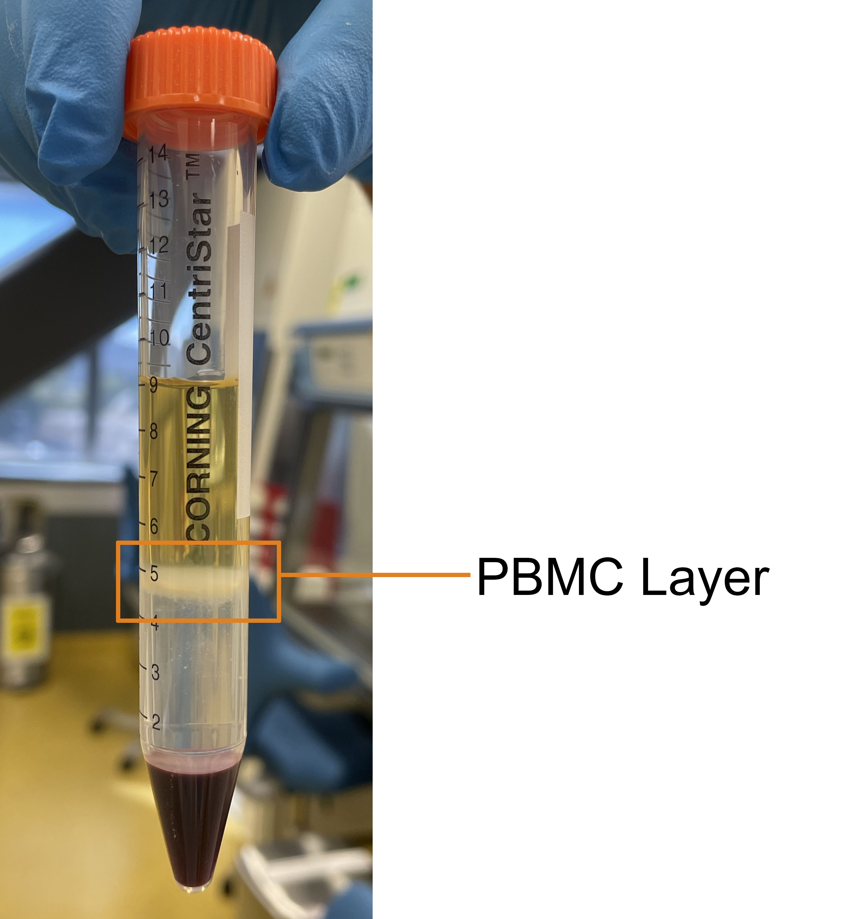
The tubes were then spun in the centrifuge for around 40 minutes which separates cell types and plasma in the sample.
Image of tube post centrifugation showing layers of separated blood. The PBMC layer is highlighted as a thin section between yellow plasma and clear separation medium.
Following centrifugation, the PBMC layer was removed and washed resulting in a small pellet of PBMC cells.
PBMC collection and resulting cell pellet.
The pellet was resuspended in media and a cell count performed to determine the number of viable cells.
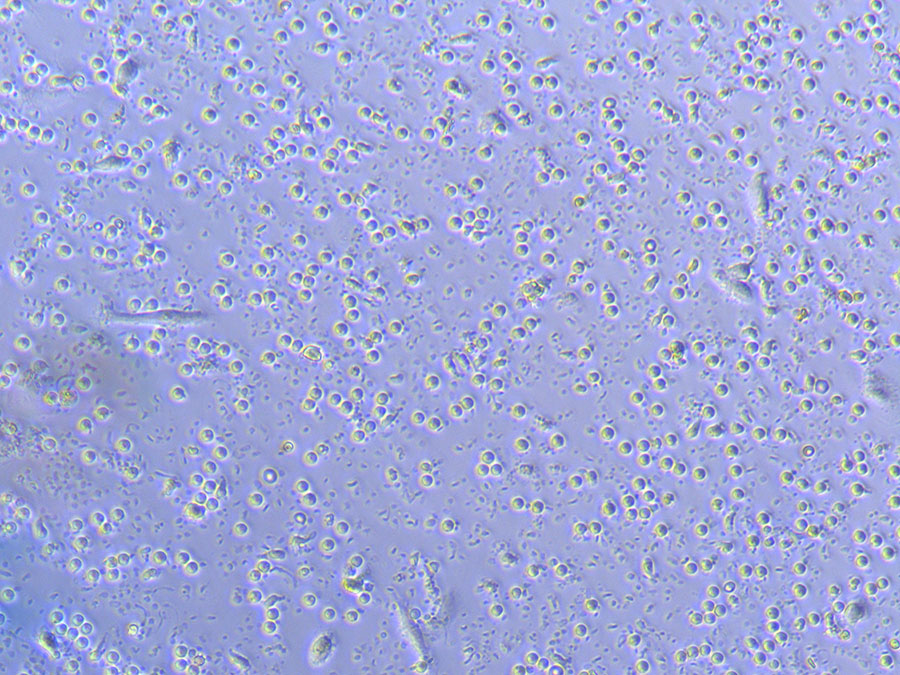
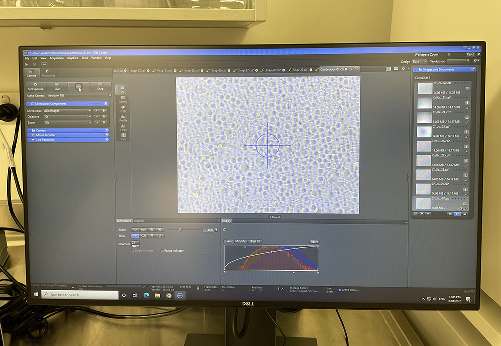
Image of densely packed PBMCs viewed under the light microscope.
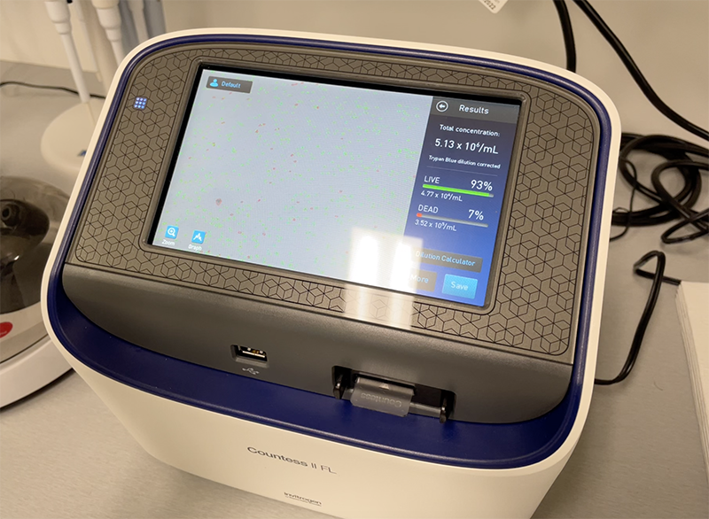
Following a cell count (using the automatic cell counter), there were around 5 million cells per mL in suspension.
Cell count showing cell count including live/dead cell percentage
Ash advised that we only need 500, 000 cells for the reprogramming (so around 100μl).
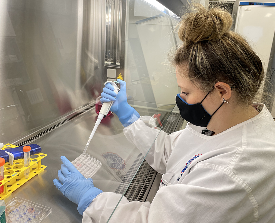
As such, we added100μl cell mix to 0.5mL media a well in a 48 culture plate.
Transferring PBMCs to 48 well culture plate
The remaining cells were resuspended in 2mL freeze media, aliquoted into two cryo-vessels (1mL each), and placed in a Mr Frosty Box (freezing container) in the – 70 freezer. They need to be transferred to liquid nitrogen in 24 hours. These frozen cell stocks operate as quality control and as a backup in case there is an issue with the reprogramming protocol (or lab mishap). Fingers crossed for a smooth journey to iPSC babies.