
After spending most of the day in the lab staining up the cut glass dishes and vessels…
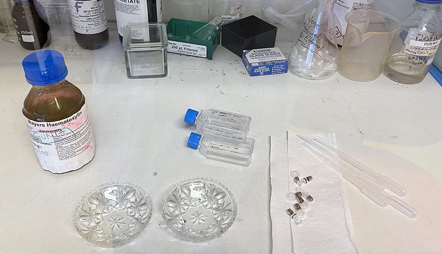
… I have emerged victorious-less.
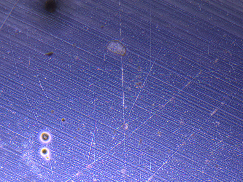
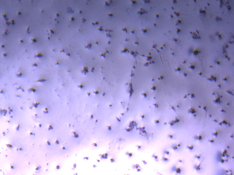
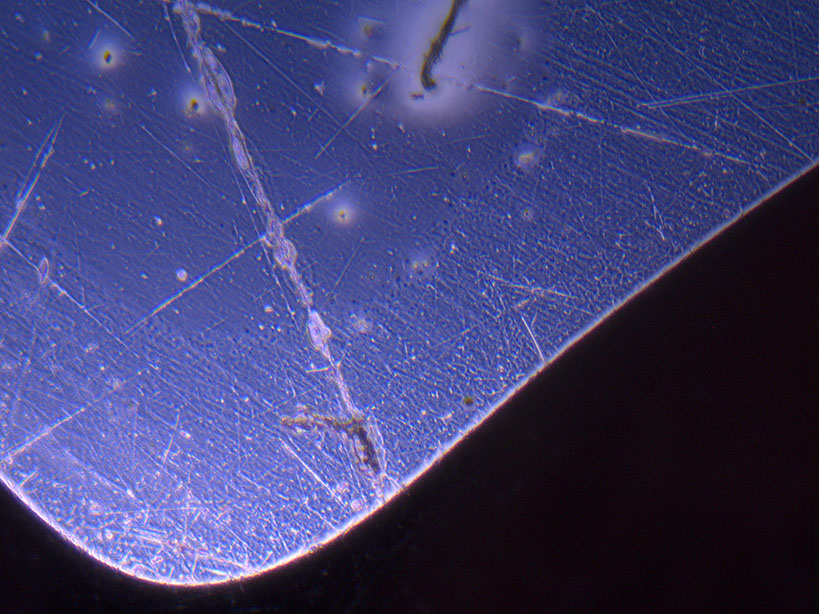 Microscope images of cut glass dishes after H&E staining on 20/01/22 showing scratches on glass surface and no cells.
There are no cells visible at all – just scratches on the surface of the glass. This is likely due to the very limited number of remaining cells which may have been further dislodged during the washing process.
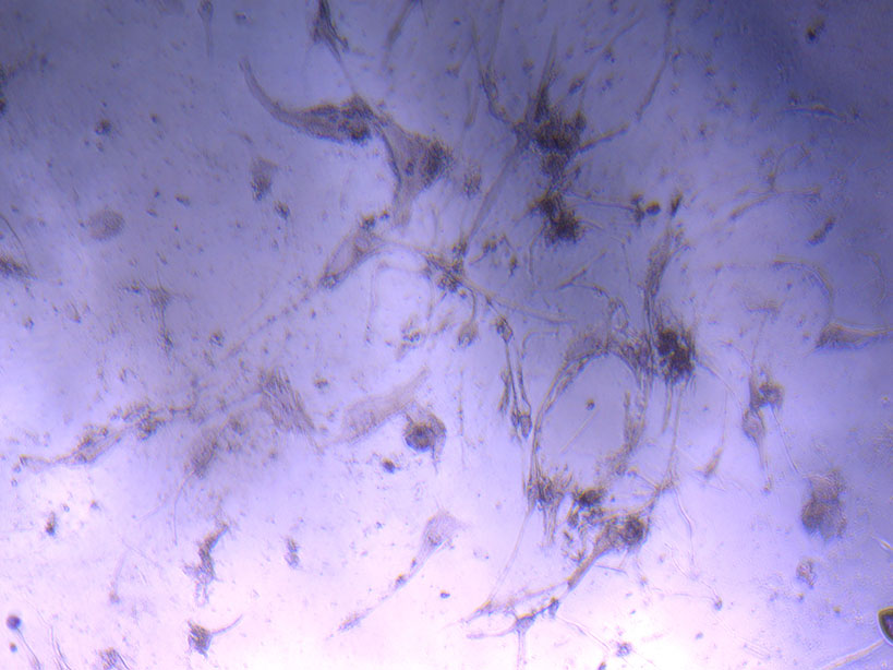
The glass vials have not fared much better. While there are cells visible, most of them are dead or dried out as it was tricky working with the small opening and 3D surface area.
 Microscope image of glass vial after H&E staining on 20/01/22. The image shows a vast number of dead cells that were not fixed in a live state.
 Microscope image of glass vial after H&E staining on 20/01/22. The image shows dried cell remnants.
The flasks show relatively good fixation of the cells, but the staining is not really visible under the light microscope in the ‘dirty’ lab.
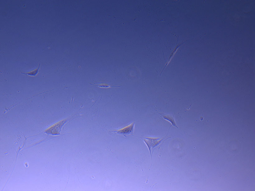
Microscope image of fixed PHGL Tumour Baby Cells after H&E staining on 20/01/22. The image shows intact fixed cells with very limited evidence of H&E stain.
I think, I will stick to Petri Dishes for the next test as they offer a more consistent surface area to work with.