
Observing the cells on 13/4/22 (24 hours after the transduction), Ash was confident that the virus had worked. While not scientifically verifiable, one of the potential indicators for success is the difference in size between standard PBMCs and ‘bloated’ PBMCs that suggest the cells have taken up the virus.
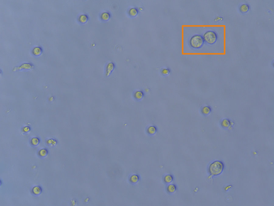
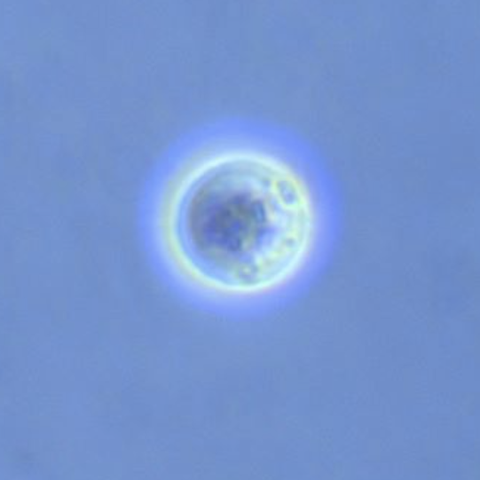
In addition to being larger, cells that may have taken on a viral load tend to have a speckled appearance. The bright halo also indicates that the cell is alive.
We transferred the cells to 2 x 60mm Petri Dishes coated with Matrigel. At this point, we just need to top up the cells with fresh media and wait (and hope) for transformation and attachment. We can also refer to the cells as RP (reprogrammed cells) rather than PBMCs.