
Following more detailed review of cell flasks, there are some hardy ‘survivors’ of my lab-free holiday.
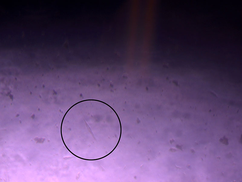 Light microscope image of cut glass dish with media containing dead cell debris and evidence of a small number of surviving cells.
After removing the old media, it was easier to see the remaining cells:
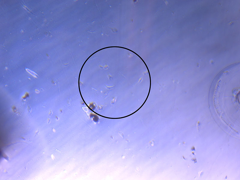
Light microscope image of cut glass dish in PBS showing evidence of a small number of surviving cells.
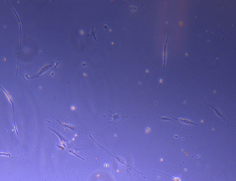
This image shows even more evidence of cell survival:
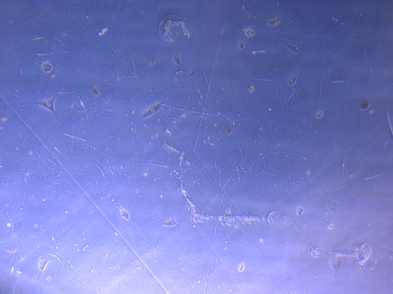 Light microscope image of cut glass dish in PBS showing further evidence of a small number of surviving cells.
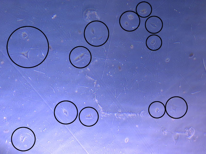 Light microscope image of cut glass dish in PBS with likely cells circled. There are a few additional potential cells visible, but I have only circled the most obvious.
To get an even better sense of survivors, I will fix (in 4% PFA) and H&E stain 2/3 of the cut glass dishes. The flatter cut glass dish and Petri dish (with more potential for cell survival will be maintained in the incubator to see how they fare over the next week).
 Light microscope image of Petri dish with fresh complete media with live cells.
I will also fix and stain the glass vessels. It is less likely that these will yield anything interesting, but it will help me troubleshoot how to do the protocol with the tiny openings – it is very difficult to effectively remove the media – even with a 20ul pipette and tip 🙁