Tag Archives: lab

Lab Training

Today, I undertook my first day of hands-on lab training. To reduce workload for my kind hosts, I tagged along with 3rd year B/Med students. As part of a unit, they are doing a study into the effects of EGCG (Epigallocatechin Gallate) – an active ingredient in Green Tea which may have positive impacts on stroke patients.  For their study, they are testing the EGCG with HBVP pericyte cells.  The protocols covered today include: cell passage, cell counting and preparing 96-well plates with consistent cell numbers.

It was good to get back into the lab and go over the basics again.  Here is a short summary of the basic protocols involved in cell culture (with some additional insights from Jo-Maree in italics).

CELL SUSPENSION

  1. Put on gloves and ensure they cover the sleeve gap of the lab coat. [Gloves are required for working in the lab. Lab glasses are required for general lab areas but are not mandatory when working in a laminar flow cabinet as the glass offers protection].
  2. Turn on UV light in laminar flow hood (10 mins) to sterilise the interior working area. [Some hood have an automatic turn/off function that enables users to easily sterilise after use. Because these hoods are manual, group users tend to sterilise before use]
  3. Turn off UV and turn on light and blower to start the sterile airflow.
  4. Organise equipment, consumables and media (1 x fluid waste vessel, 1 x pipette waste vessel, pipette and pipette gun, pericyte media, PBS– and TrypLE . Spray all items (including gloved hands) with 70% Ethanol before placing in hood. [Do not place cells in front of waste vessels as this increases contamination due to laminar flow.  PBS– refers to phosphate buffered saline. The — is shorthand for PBS without Calcium (Ca2+) and magnesium (Mg2+) . These minerals help cells adhere to the surface of the culture flask. As such PBS — (without Ca2+ and Mg2+) is used when passaging cells (splitting them into additional flasks).  PBS++ (with Ca2+ and Mg2+) is used to promote cell adhesion. TrypLE is a form of trypsin (a digestive enzyme that detaches cells from adhering to the flask surface). TrypLE is milder and works well with HBVP cells.]
  5. Aliquot media from shared stock and place in heating bed. [The lab has a metal heating bed instead of a water bath. This is a new development and was implemented to reduce the likelihood of contamination as water baths (37 degrees, warm and moist) are perfect breeding grounds for bacteria and a known source of potential  contamination in cell culture).
    Metal Lab Beads for media heating
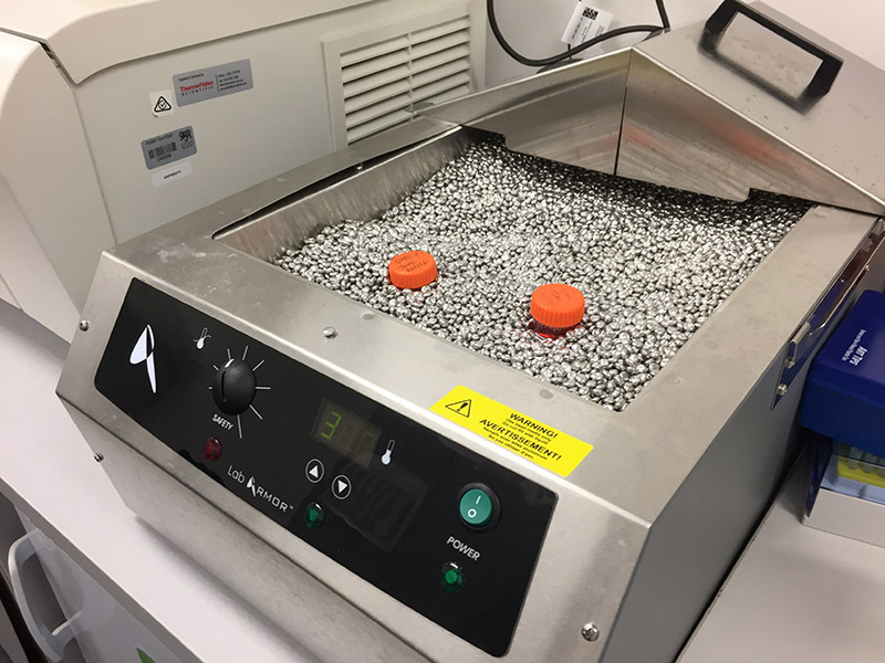
‘Water bath’ using metal pellets to warm media to 37°C (body temperature) – very cool
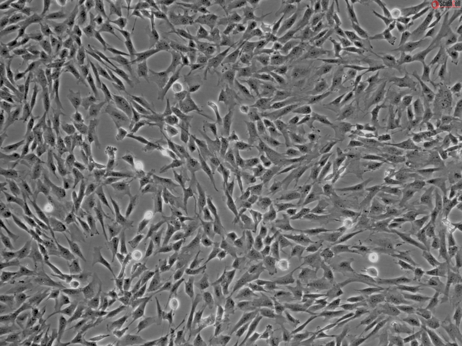
  6. Collect cells from incubator and view under the microscope. Check cells for any contamination or odd changes in morphology.  [The HBVP cells used today were plated on Friday and are at passage 4 (P4) meaning that they have been split 4 times (although full passage numbers are not disclosed by cell suppliers). Today they were approx. 80% confluent (i.e. they have filled approx. 80% of the available space on the surface of the flask). It is important to not let cultures grow to 100%, as this impacts on cell growth and behaviour. Similarly, cells should be maintained at 30 – 40% min as too few cells also impairs growth. The lab also tends to use cells up to P8 or P9 only to ensure reliable results.]

    Microscope
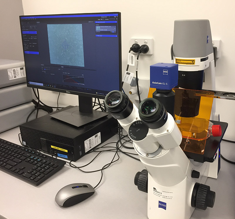
Microscope connected to digital capture

    Cells observed under the light microscope
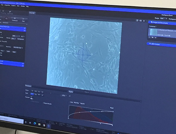
    Microscope view of cells displayed on the computer screen

  7.   Review cells for any contamination or odd changes in morphology.
  8. Spray flask with 70% ethanol and place into laminar flow hood.
  9. Remove nutrient medium [This can be poured off into the liquid waste vessel, if following correct technique.]
  10. Add 5mL PBS– to culture flask using 5mL pipette.
  11. Gently rotate the flask to wash the cells.
  12. Remove (pour off) PBS solution.
  13. Add 5mL TrypLE (trypsin) and place flask in incubator for 5 min.
  14. After 5 min, check that cells have detached.
  15. Add 10mL complete nutrient medium. [The nutrient medium contains fetal bovine serum FBS which neutralises the trypsin and stops cell digestion]. 
  16. Add cell solution to a 15mL tube.
  17. Spin cells in the centrifuge at 200G for 10 min (room temp). [It is important to add a counter balance when using the centrifuge.]
  18. Check that there is a small cell pellet in the base of the tube.
  19. Spray tube with 70% ethanol and return to laminar flow cabinet.
  20. Gently pour off liquid into the liquid waste vessel.
  21. Resuspend cells in 2mL complete nutrient medium.

CELL COUNTING

The cells are now ready for counting using a traditional haemocytometer – a glass slide with two chambers with a grid of 9 x 1mm squares used to count cells.

  1. Set up haemocytometer by adding the coverslip. [It helps to breath on the slip to create a good seal]
  2. Remove 20 μL of cell suspension.  Remove from hood. [You can place this on a piece of parafilm as it does not need to remain sterile.]
  3. Add 20 μL Trypan Blue to the 20 μL cells and pipette gently to mix the two solutions.  [Trypan Blue stains dead cells as the dye can penetrate through the dead cell membrane. This is used as a mechanism to help count viable cells only].  
  4. Add 10 μL to each chamber.
  5. Since HBVP cells are relatively large, count the cells in the large four corner squares. [When counting cells have a system to avoid doubling up on cells i.e. count top and right only of overlapping cells. Also check to make sure cells are evenly dispersed. If they are clumped together, you will need to suspend cells more thoroughly – i.e. mix with pipette]
  6. Add all cell counts from the corner squares together: 127 + 104 + 116 + 133 = 480 ÷ 4 (number of squares to get an average) = 120 x 104 x 2 (due to dilution with Trypan Blue) = 480 x 104. This is 4.8 x 106 (so 4.8 million cells in the 2mL suspension).
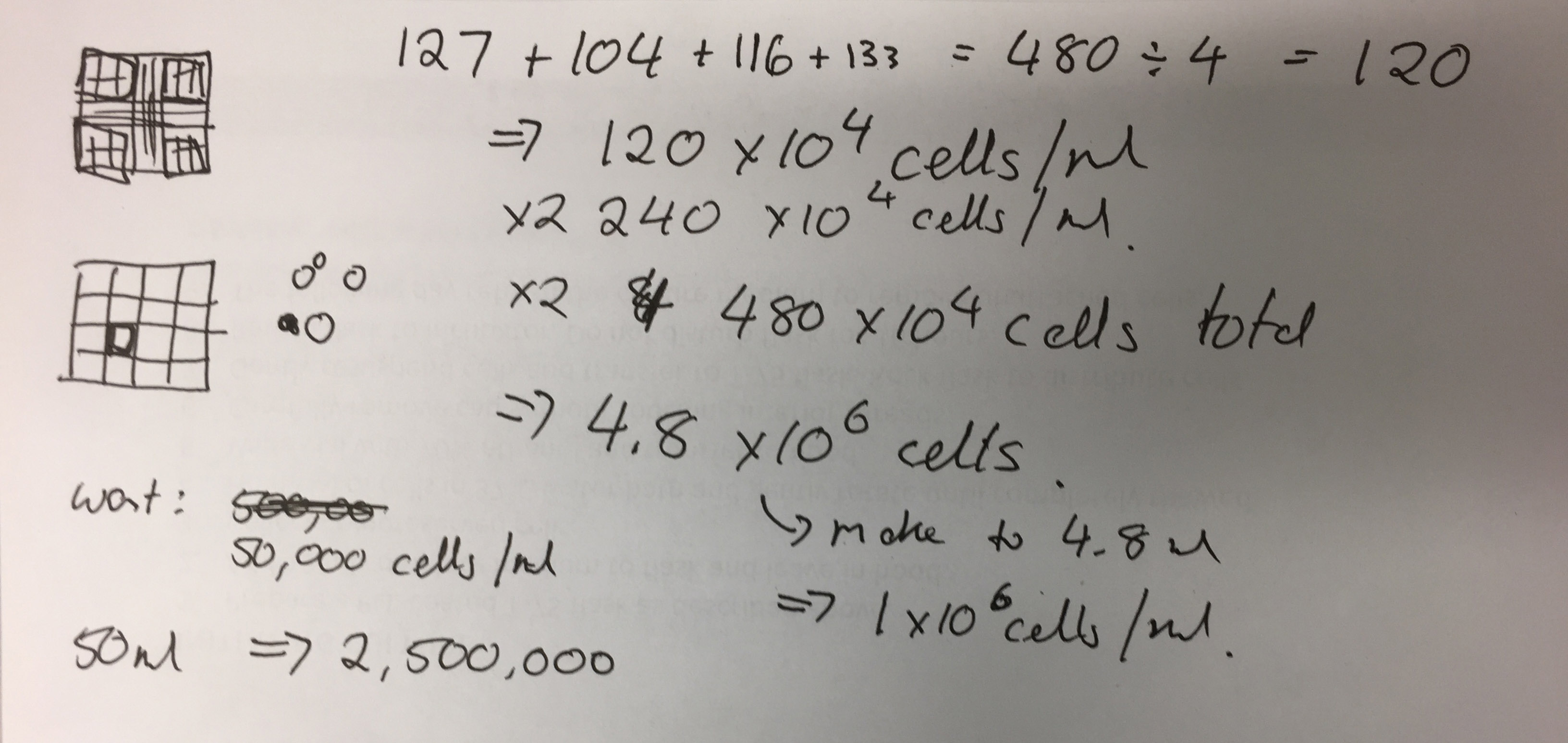
    Cell Counting
    Photo of Jo-Maree’s calculations
  7. To make it to 1x 10⁶ add 280μL to the 200μL.

The students set up 96 well plates with 100μL @ 5000 cells per well  as part their protocol.

CELL PASSAGE

  1. Add 1mL of the 1x 10⁶ cell suspension to a new flask.  Add 9mL fresh warm complete medium and gently rotate the flask to disperse cells.
  2. Remove cells from hood and place them in the incubator. [The cells will be confluent within 48 hours]. 
  3. Remove media, used pipettes and waste vessels from the hood.
  4. Place pipettes and pipette tips in biohazard bins. [If bins are full, take to dirty autoclave station and loosely secure bag with autoclave tape.]
  5. Add liquid waste to bleach to kill any remaining cells. [After 24 hours, all organisms are destroyed and the remaining liquid can be safely disposed of.]
  6. Place waste vessels in bleach bucket in sink area. [After 24 hours, these can be washed and left to dry for re-use].

ON A SIDE NOTE:

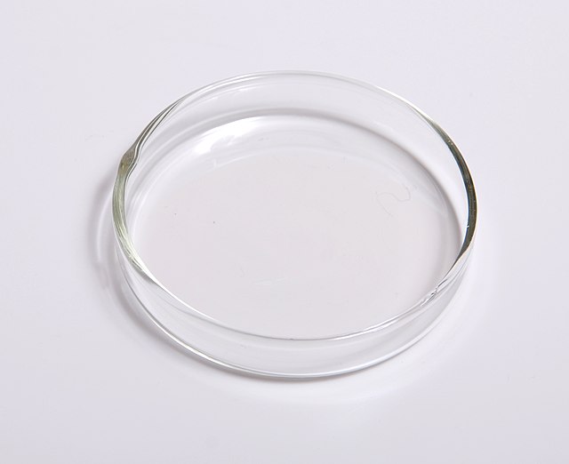
Poly-L-lysine is the correct coating to promote cell adhesion on glass surfaces. Jo-Maree has some in stock, so I am all set to work with my Petri dishes as part o the initial cell culture training.

Material supplies: Glass Petri Dishes

My primary lab contact is currently on leave so I am using the time to identify materials for experimentation. I am keen to grow and stain my cells on diverse materials (glass, porous and non-porous scaffolds etc). An easy start is to use glass Petri dishes with different coatings to encourage cell adherence. The use of coatings may also enable me to encourage cells to grow in particular patterns.

I’ve done a bit of searching via Researchgate and it seems that common surface coatings to encourage cell attachment to glass include:

  • Hyaluronic acid
  • Poly-D-Lysine – my current choice
  • Fetal Calf Serum
  • Bovine serum albumin
  • Gelatin
  • Fibronectin
  • Laminin
  • Collagen

I will ask for advice at the next lab meeting.

There were also suggestions to etch the glass surface with concentrated nitric acid and then wash and autoclave. At the UTAS School of Creative Arts and Media, we are fortunate to have access to a glass cutter and laser engraver. So instead of using acid, I will use the laser to score a design into the base of a large 150mm Petri dish.

Glass Petri Dish

Glass Petri dish by Lilly M via Wikimedia

Luckily Petri dishes are easy to purchase online. I just need to make sure the dishes are suitable for autoclave sterilisation (e.g.  borosilicate glass rather than soda lime glass).

Training with HBVP [Human Brain Vascular Pericytes]

Despite UTAS improving the turn-around time for ethics clearance applications, it will take some time for the new application to be processed. Of course, we do not want to be idle, so the time pending formal approval will be used for project training.

While I have worked with cell and tissue culture previously as part of my PhD study at QUT, it has been over five years since I have actively worked in a lab environment. As such, it is important that I undertake training to ensure I am up to date with protocols and understand the working methods used at the UTAS School of Medicine.

To get up to speed, Brad and Jo-Maree suggested that I start working with HBVP Human Brain Vascular Pericyte cells.

HBVP Cells - via ScienCell

100 x phase contrast microscope image of HBVP cells via ScienCell.

These cells are a commercially available cell line and routinely used by the group for stroke research. The advantage of working with commercially available cell lines is that they have established protocols for optimum culture. As long as the research group is authorised to work with cell lines, they also do not require additional ethical clearance.

Culturing HBVP cells will not only enable me to brush up on cell culture techniques, they will also enable me to explore the behavioural characteristic of a different cell type. Indeed, Brad informed me that HBVP cells grow over microvessels and at later passage numbers, tend to form circular formations in culture.

whiteboard

Whiteboard drawing from meeting showing circular cell formation of HBVP cells and set ups for co-culturing cells via chamber inserts and microfluidics.

Brad and his group have been keen to work with 3D vascular scaffolds as part of their research, so the project and training stage provides an opportunity to work with some of Dietmar’s biofabricated tubes from the QUT Centre for Regenerative Medicine. After all, it is always better if training can also be productive!

Project Meeting

We had a detailed project planning and ethics clearance review meeting on Monday. It was heartening to hear that the ethics clearance documentation is almost ready for formal submission.

These are some of the processes we are expecting to undertake:

  • Cell culture of fibroid cells (optimisation of culture methods for 2D and 3D environments, cellular response and proliferation testing)
  • Co-culture of fibroid cells with other cell lines
  • Genetic profiling of primary fibroid cells
  • Immortalisation of primary fibroid cells via established commercial kit (e.g. Applied Biological Materials (ABM) cell immortalisation kits)
  • Reprogramming of primary cells to generate induced pluripotent stem cells (iPSCs) via established commercial kit (e.g. Epi5™ Episomal iPSC Reprogramming Kit available via Thermo Fisher)
  • Cell culture of reprogrammed or immortalised fibroid cells (optimisation of culture methods for 2D and 3D environments, cellular response and proliferation testing)
  • Genetic profiling of reprogrammed or immortalised fibroid cells and cells lines (if successful)
  • Fixing and staining of cells
  • Light and confocal microscopy of cultured cells
  • Timelapse video of cultured cells

Other potential processes include:

  • Green Fluorescent Protein (GFP) cell tagging [UTAS]
  • Scanning Electron Microscopy (SEM) of cultured cells [UTAS]
  • Transmission Electron Microscopy (TEM) of cultured cells [UTAS]
  • Histopathology of cultured 3D structures [UTAS or QUT]
  • Development of Gastruloids, Organoids or Neurospheres (self-organised 3D cell masses) [UTAS]

I was delighted to hear that my desires for creating aggregates of cells via the production of gastruloids or organoids was not outside the domain of possibility. Jo-Maree has produced neurospheres (balls of neural stem cells) previously, so there may be scope (and hopefully time) to experiment with cell clusters.

Gastuloids are of particular interest to me as they are cell clusters that display features of early embryo development. Staining and fluorescence imaging (apart from being visually stunning) enables the visualisation of tissue organisation as shown in this figure and caption from the Nature publication ‘Multi-axial self-organization properties of mouse embryonic stem cells into gastruloids’:

Tissue organisation in gastruloids

Tissue organization in gastruloids a, Gastruloids formed from Sox1GFP;BramCherry (SBR) line and stained for Sox2 expression (Sox1GFP and SOX2 signals are displayed in green and magenta, respectively). White arrowheads indicate tubular SOX2/Sox1-positive neural structures. Red arrowheads point to the presumptive digestive tube. b, WISH on 8-µm transverse cryosections of gastruloids at 144 h AA using Sox2 and Meox1 antisense probes, counter-stained with Nuclear Fast Red. Sox2-positive cells localized predominantly in a compact dorsal domain, whereas Meox1 signals were found in two bilateral domains. The domain of expression of each gene is outlined with white dashed lines. c, Haematoxylin and eosin staining of transverse paraffin sections of different gastruloids at 120 h AA, showing the diversity of cell types and several levels of tissue organization. d, Gastruloids formed from Sox1GFP;BramCherry ESCs were fixed and stained at 168 h AA for OLIG2 (top, white), PAX3 (middle, red) and PAX7 (bottom, red). Scale bars as indicated. c, d, Gastruloids formed from Sox1GFP; BramCherry ESCs collected at 168 h AA and stained for SOX17 (magenta, c) or CDX2 (magenta, d). Scale bars as indicated.

Gastruloids are created from embryonic stem cells, although iPSC cells (cells that have been reprogrammed into a stem cell like state) have also been used. As such, if I have success with reprogramming my fibroid cells to iPSC cells, I could use them to make, and learn more about, gastruloids.

During our discussion, we decided it would be a good idea to include an optional alternative to the fibroid cells – just in case there is an issue with contamination or the freezing/thawing process. While I have isolated skin cells (fibroblasts) from hair follicles and skin grafts previously as part of the HSE Project at QUT, we decided on skin scrapings.   This approach was selected as will enable the isolation of skin cells, is not too invasive and is well established within the School of Medicine.

Once I’ve included this information including the protocol, I should be ready to submit the final ethics clearance document.

Beccari, L., Moris, N., Girgin, M., Turner, D.A., Baillie-Johnson, P., Cossy, A.C., Lutolf, M.P., Duboule, D. and Arias, A.M., 2018. Multi-axial self-organization properties of mouse embryonic stem cells into gastruloids. Nature562(7726), pp.272-276.

Lab Meetings

One of my favourite things about being integrated in a research lab environment is that most research groups have regular meetings that give insight into research progress. Not only do these meetings allow members to gain insight into the various projects being undertaken within the group, they also enable troubleshooting and critical feedback.

Meeting NotesNow that I am officially part of the Stroke Group (with Synapse collaborators A/Prof Brad Sutherland and Dr Jo-Maree Courtney), I attended my first Tuesday afternoon meeting. Brad was the presenting speaker who shared some of his research from a Postdoc undertaken at the University of Oxford some years ago. The talk focused on “Glutathione  and its response following stroke”. While some of the discussion (particularly in relation to the Glutathione  pathway) was beyond my knowledge, it was still useful to consider the discussion in terms of experimental design.  It was also great to be reminded that sometimes outcomes are unexpected or unsatisfying, as they may contradict expectations.

Step 1: Lab Inductions

Over the past few weeks I’ve been preparing for my exciting foray back into a lab environment. I am hopeful that it is ‘just like riding a bike’, although my finger dexterity for working one handed to open media bottles will likely need some renewed practice.

To date, I have completed my online training to ensure I am familiar with laboratory rules. At UTAS there are three levels Green (“general safety standards”), Amber (“higher level of risks and hazards associated with the space and activity”) and Red (“highly specialised inductions associated with an activity”). For access into a Physical Containment Level 2 (PC2) lab environment for cell culture, I am required to complete the inductions for all levels including Red Level Chemical and PC2 training. While I am still familiar with much of this information from my time at QUT, it is a good reminder of safety regulations. Even though it is a requirement, as an artist and scientific outsider, I feel it is even more important to set a strong example and adhere to all institutional guidelines and best-practice exemplars.

On Tuesday 29/6, I had my site induction with the lab technician Alex. Like many research labs, the work areas are like a maze and it will take me a bit of time to familiarise myself with the various locations for microscopy, bench work and cell culture. I am excited to be in the ‘Dirty’ Biobanking area where I will work with the rather excellent Dr Jo-Maree Courtney on culturing and hopefully immortalising my Tumour Baby (Fibroid) cells. Note that the term ‘dirty’ does not reflect my hygiene standards, but rather relates to the status of the cells as primary cells that have not undergone testing to ensure they are free from pathogens including potentially lab-borne infections such as mycoplasma.

PC2

As you can see from this photo of the PC2 entry, there are strict lab rules to ensure safety. Photography (without permission) is not permitted, although I have approval form the lab manager to document my work over the residency period. As a visual person, I find that documenting protocols via photos works well for me and helps facilitate memory.


As part of the residency, I am also challenging myself to use drawing as a mode of capturing my engagement. This approach was inspired by a recent drawing workshop ‘Drawing on the Brain’ I undertook with Dr Megan Walch at the Moonah Art Centre. She reminded me that drawing is really effective tool for memory and building neural pathways in relation to experience.  I’ll have to see if I can maintain the process..

Post-holiday update

I have returned from the festive season break and started back in the lab.

Let’s start with the good news! There is no visible infection in any of the vessels including cut glass dishes and vials. My flasks are doing OK and there are cells actively growing (despite evidence of cell death indicated by cell debris).

Now for the bad news…

There has been mass death. Despite the slow growth rates of my cells, 3 weeks is just too long to leave cells starving and without ongoing maintenance. That said, there is evidence (in cell debris) that a good number of cells did grow in the vessels during my absence. This shows that the overall plan should work.

The plan for today is:

  1. Make up new media and FBS aliquots.
  2. Feed cells (i.e. replace media with fresh solution)
  3. Remove dead cells from all cut glass dishes.
  4. Collect dead cells via centrifugation and fix in PFA.
  5. Fix cells in some of the older flasks, fix in PFA and stain with H&E.

If I have time, I will also:

  1. Bleach and wash glassware and prepare for autoclaving.
  2. Autoclave glassware.